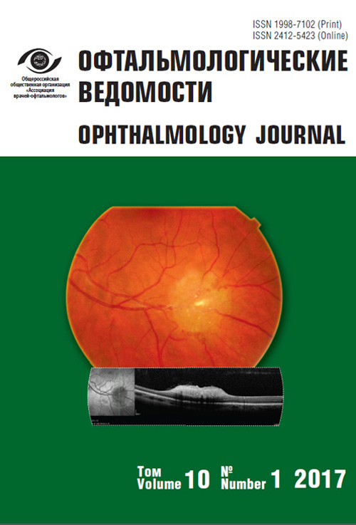
Retinal astrocytic hamartoma in tuberous sclerosis
- Authors: Astakhov Y.S.1, Nechiporenko P.A.1, Atlasova L.K.1, Titarenko A.I.1, Monakhov K.N.1, Romanova O.L.1, Molodykh K.Y.1
-
Affiliations:
- I.P. Pavlov First St Petersburg State Medical University
- Issue: Vol 10, No 1 (2017)
- Pages: 97-101
- Section: Articles
- URL: https://journals.eco-vector.com/ov/article/view/6322
- DOI: https://doi.org/10.17816/OV10197-101
- ID: 6322
Cite item
Abstract
The article describes a clinical case of long-term undiagnosed tuberous sclerosis with retinal astrocytic hamartoma. Etiology, pathogenesis, clinical and pathomorphological diagnostic criteria of tuberous sclerosis are also described.
Keywords
Full Text
Tuberous sclerosis (TS), also known as Morbus Pringle-Bourneville, or adenoma sebaceum, is a genetic neuroectodermal disorder affecting internal organs, bones, eyes, skin, and nervous system, with the formation of benign tumors called hamartomas [2, 3].
The disease was first described in 1862–1863 by Frederich Daniel von Recklinghausen, who detected multiple heart tumors and brain changes at during an autopsy of a baby. In 1880, Desire-Magloire Bourneville, a French neurologist, provided a detailed description of the neurological and pathological changes in the brain of a girl with convulsive paroxysms, mental deficiency, and skin disorders. In 1890, John James Pringle, a British dermatologist, first described skin manifestations on the faces of patients with TS and used the term “congenital adenoma sebaceum.” Since then, the disease is called Bourneville-Pringle disease. Later, many authors stressed that TS is a multisystem disorder characterized by multiple lesions across different organs. In 1908, Н.Vogt outlined the classic clinical triad of the TS complex (TSC): sebaceous gland adenoma, epileptic seizures, and mental deficiency. In 1920, Jan van der Hoeven, an ophthalmologist, described specific changes in the ocular fundus which were typical of TS, i.e., a mulberry lesion or phacoma, later termed phacomatosis [1–3].
TS is inherited in an autosomal dominant pattern; it is caused due to de novo mutations in approximately 80% of patients. Two genes are involved in TS development: TSC1 (located on the long arm of chromosome 9 at position 34) which encodes hamartin and is responsible for type 1 TS, and TSC2 (located on the short arm of chromosome 16 at position 13) which encodes tuberin and is responsible for type 2 TS. The disease is characterized by varying gene expression and almost 100% penetrance. TC is estimated to be prevalent in one per 30,000 individuals; the prevalence varies between 1:6000 and 1:10,000 in newborns. There is no gender predisposition to the disease [3, 4].
Diagnostic criteria for TS were developed in 1998, which state that the diagnosis can be established if a patient has at least two primary symptoms or one primary and two secondary symptoms, probable (possible) diagnosis if a patient has one primary and secondary symptom each, and suspected (questionable) diagnosis if a patient has one primary or at least two secondary symptoms (Table 1) [2, 3, 5].
Table 1. Diagnostic criteria of tuberous sclerosis
Таблица 1. Диагностические критерии туберозного склероза
Primary signs | Secondary signs |
Angiofibromas and fibrous plaques on the face | Multiple recesses in tooth enamel |
Nontraumatic periungual fibroma | Hamartomatous rectal polyps |
Hypopigmented areas (>3) | Bone cysts |
Shagreen patch | Cerebral white-matter migration tracts |
Retinal hamartomas | Gingival fibromas |
Cortical tubers | Hamartomas in the internal organs |
Subependymal nodes | Retinal achromic patch |
Giant cell astrocytoma | Confetti hypopigmentation |
Cardiac rhabdomyomas | Multiple renal cysts |
Lymphangioleiomyomatosis in lungs | |
Multiple renal angiomyolipomas |
Ocular manifestations of TS usually include retinal or optic nerve hamartomas [1–3]. There are two main clinical types of retinal astrocytic hamartomas: small, noncalcified, flat, smooth tumors appearing as mild thickening of nerve fibers; and large, calcified, whitish-yellow, knotty tumors (mulberry lesions). The same tumor may include areas of both types due to gradual calcification.
Differential diagnosis of TS includes retinoblastoma, myelinated nerve fibers, and retinal granuloma; in case of optic nerve harmatoma, the diagnosis should also include optic disc drusen (astrocytic hamartomas on the optic nerve occasionally termed giant drusen of the optic nerve) and papillitis (optic disc edema in patients with TS caused due to intracranial hypertension).
Retinal astrocytic hamartomas are usually asymptomatic and do not require treatment. However, in case of rare complications, such as vitreous or retinal hemorrhage, spread of the tumor to the vitreous body, and retinal detachment, a symptomatic therapy is recommended. Patients and their family members should be examined to identify TS symptoms.
Approximately 50%–90% of patients have skin lesions on the face that commonly appear in childhood and manifest as multiple, tumor-like, yellowish or brownish, smooth-surfaced formations of up to 0.5 cm in diameter with telangiectasias. Sub- and periungual fibromas are observed in 17%–52%, hypomelanotic spots in 90%, coffee-with-milk-colored spots in 15% and shagreen patches in 20%–68% of patients. Hair depigmentation and fibrous growth of the oral mucosa are very rare [1, 5, 6]. Neuropsychiatric manifestations include epilepsy in 80%–90% and impaired intellectual function in 48% of patients, which are probably induced by the growth of glial structures in the brain [1]. Cardiovascular manifestations include disorders of cardiac conduction and rhythm that may result in cardiac arrest due to cardiac rhabdomyomas in 30%–60% of patients [1]. Approximately 10% of patients develop hepatic angiomyolipomas and hamartomas; 50%–78% show rectal polyps and 47%–85% show bilateral angiomyolipomas and multiple cysts in kidneys [1].
Clinical case description
The patient was initially admitted to the Dermatology Department of the Pavlov First Saint Petersburg State Medical University and then referred to an ophthalmologist for consultation. The 35-year-old man complained of painless non-itchy rash on the face, upper body, fingers, and extensor surfaces of the upper and lower extremities. The disease onset occurred at the age of two years, when the patient first manifested with spotty rash on the face. Skin lesions on the cheeks were assumed to be caused by lactose intolerance. The patient received no treatment, but elimination of lactose from the diet was recommended. Later, small asymptomatic nodular lesions emerged around the nasolabial folds; a spotty rash appeared on the back and lower extremities. At the age of five years, the patient manifested with epileptic seizures that persisted until the age of 10 years (frequency unknown).
In 1995, the patient first noticed an itchy elbow rash that was considered as psoriasis by a local dermatologist. The disease was relatively mild with occasional exacerbations. However, small nodular rashes continued to appear on the face.
In 2012, the patient presented with rash on the face, neck, back, hands, and the lower limbs. He was diagnosed with psoriasis and was put on a specific treatment. The psoriatic rash regressed in response to the therapy; however, the rash on the face remained unchanged. In 2015, he was referred to a dermatology clinic, where TS was first suspected, and in-patient examination and treatment were recommended.
Upon examination, the patient’s condition was found satisfactory; he had clear consciousness with some dormancy. He had a widespread rash on the face, neck, scalp, left eyebrow, right shoulder, back, and periungual folds. The rash, which was present on the nose wings, nasolabial folds, cheeks (butterfly pattern), and chin, was symmetrical, dense, confluent, red, noninflammatory, and of papular type, with a diameter of 1–3 mm. Papules were hemispherical, with sharp limited outlines, dense consistency, and smooth surface (Figure 1). Slightly raised papular elements were found on the gingival mucosa. Pathomorphological examination of the papular elements on the nose wings revealed an expansion of the sebaceous glands and collagen fibers and vasodilatation. The patient was diagnosed with angiofibroma accompanied with signs of inflammation.
Fig. 1. Multiple facial skin fibromas
Ophthalmological examination revealed normal visual function of both eyes and no pathological changes in the anterior segment. A large yellowish lesion, with twice the diameter of the optic disc, protruding into the vitreous and characterized by clear outlines was detected on the fundus of the left eye along the inferotemporal vascular arcade close to the macular area (Figure 2). B-scan ultrasonography revealed an elevated area on the internal eye tunic with an inhomogeneous retinal counter. Optical coherence tomography allowed visualizing a moderately hyperreflective homogeneous formation at the level of internal neuroepithelium with separate inclusions (calcificates). Ultimately, the patient was diagnosed with a TS-related retinal astrocytic hamartoma of the left eye.
Fig. 2. Fundus photography: large yellowish clearly outlined prominating lesion
Fig. 3. Optical coherence tomography: moderately hyperreflective homogeneous lesion at the level of the inner neuroepithelium layers with several inclusions (calcifications)
The patient showed five primary symptoms, including angiofibromas on the face and fibrous plaques on the forehead, nontraumatic periungual fibromas, hypopigmented areas, shagreen patches, and retinal astrocytic hamartoma; and three secondary symptoms of TS, including gingival fibromas, confetti hypopigmentation, and renal cyst Disease history regarding epileptic seizures and skin rash in the early childhood also helped to understand the nature of the disease. Eventually, the patient was diagnosed with TS complicated by limited psoriasis.
This clinical case had two specific features: 1) late diagnosis of TS, and 2) unusual combination of skin manifestations of TS with psoriasis, which may have hampered the diagnosis. Practicing physicians may be less familiar with the clinical features of TS. Variable symptoms, their manifestations at different periods of life and combination with psoriasis observed in this case, demonstrate the difficulties associated with the diagnosis of TS. Nevertheless, careful collection of history data and systematic analysis of specific clinical features will ensure timely identification of the disease. Examination of the ocular fundus by an ophthalmologist and optical coherence tomography of the retina will facilitate the diagnosis in such cases.
About the authors
Yury S. Astakhov
I.P. Pavlov First St Petersburg State Medical University
Author for correspondence.
Email: astakhov73@mail.ru
MD, DMedSc, professor. Ophthalmology Department.
Russian Federation, Saint PetersburgPavel A. Nechiporenko
I.P. Pavlov First St Petersburg State Medical University
Email: glaz@doctor.com
MD, PhD, assistant. Ophthalmology Department
Russian Federation, Saint PetersburgLiya K. Atlasova
I.P. Pavlov First St Petersburg State Medical University
Email: kasia66@mail.ru
MD, ophthalmologist. Ophthalmology Department
Russian Federation, Saint PetersburgAleksandra I. Titarenko
I.P. Pavlov First St Petersburg State Medical University
Email: aleksandra-titarenko@yandex.ru
MD, resident. Ophthalmology Department
Russian Federation, Saint PetersburgKonstantin N. Monakhov
I.P. Pavlov First St Petersburg State Medical University
Email: molodyhkristina@mail.ru
MD, DMedSc, professor. Dermatovenerology Department
Russian Federation, Saint PetersburgOksana L. Romanova
I.P. Pavlov First St Petersburg State Medical University
Email: molodyhkristina@mail.ru
MD, PhD, assistant professor. Dermatovenerology Department
Russian Federation, Saint PetersburgKristina Y. Molodykh
I.P. Pavlov First St Petersburg State Medical University
Email: molodyhkristina@mail.ru
resident. Dermatovenerology Department
Russian Federation, Saint PetersburgReferences
- Дорофеева М.Ю. Туберозный склероз у детей // Российский вестник перинатологии и педиатрии. – 2001. – № 4. – С. 33–41. [Dorofeeva MYu. Tuberoznyy skleroz u detey. Rossiyskiy vestnik perinatologii i pediatrii. 2001;(4):33-41. (In Russ.)]
- Мосин И.М., Дорофеева М.Ю., Балаян И.Г., Яркина О.С. Офтальмологические проявления туберозного склероза // Российский вестник перинатологии и педиатрии. – 2012. – № 5. – С. 77–81. [Mosin IM, Dorofeeva MYu, Balayan IG, Yarkina OS. Oftal’mologicheskie proyavleniya tuberoznogo skleroza. Rossiyskiy vestnik perinatologii i pediatrii. 2012;(5):77-81. (In Russ.)]
- Страхова О.С., Катышева О.В., Дорофеева М.Ю., и др. Туберозный склероз // Российский медицинский журнал. – 2004. – № 3. – С. 52–54. [Strakhova OS, Katysheva OV, Dorofeeva MYu, et al. Tuberoznyy skleroz. Rossiyskiy meditsinskiy zhurnal. 2004;(3):52-54. (In Russ.)]
- Kwiatkowski DJ, Reeve MP, Cheadle JP, Sampson JR. Molecular Genetics. In: Nuberous Sclerosis complex: from Basic Science to Clinical Phenotypes. Ed by Curatolo P. London, England: Mac Keith Press; 2003. 228-263 pp.
- Roach ES, Di Mario FJ, Kandt RS, Northrup H. Tuberous Sclerosis Consensus Conference: Recommendations for Diagnostic Evaluation. Journal of Child Neurology. 1999;14:401-407. doi: 10.1177/088307389901400610.
- Rogers RS, O’Connor WJ. Dermatologic Manifestations. In: Tuberous Sclerosis. Ed by M. Gomes, J. Sampson, V. Whittemore. New York; Oxford: Oxford University Press; 1999. 160-180 pp.
Supplementary files


