

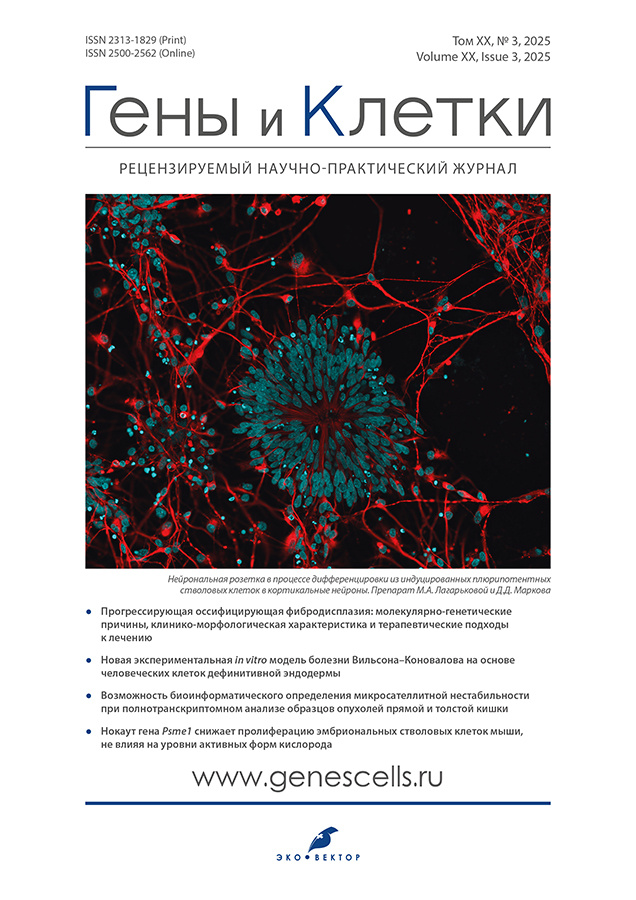
")


Genes & Cells

Peer-reviewed scientific and practical journal
Editor-in-chief
- Maria A. Lagarkova, Dr. Sci. (Biology), professor
ORCID iD: 0000-0001-9594-1134
Publisher
Founders
- Genes & Cells llc
- Lopukhin Federal Research and Clinical Center of Physical-Chemical Medicine of Federal Medical Biological Agency
- Eco-Vector
About
“Genes & Cells” (the old name is “Cell Transplantology and Tissue Engineering”) is a quarterly peer-reviewed scientific and practical journal.
The sections of the journal are formulated to fully disclose the target topics of the publication, convey to readers the opinions of leading experts in the field of biomedical technologies on topical issues of concern, acquaint them with the most significant recent foreign and domestic research, materials of thematic conferences, present analytical information on fundamental issues of biomedical technologies trends in the biotech business.
Thus, the journal does not just keep up with the time, but jointly with the website, which is an independent scientific information and analytical mass media, changes the views of representatives of medical specialties on the possibility of using biotechnologies in clinical practice; at the same time, we do not juggle with facts and do not impose subjective unverified data — all the tools of the journal and site are to convey to a wide circle of readers only objective scientific and analytical information.
Journal headings
- expert opinions
- cell technology news
- reviews
- original research
- clinical experience
- discussion and general theoretical work
- stem cell business
Types of manuscript
- reviews
- systematic reviews and metaanalyses
- original research
- clinical case reports and series
- letters to the editor
- short communications
- clinial practice guidelines
Publications
- in English and Russian
- quarterly, 4 issues per year
- continuously in Online First
- distribution in Hybrid model (subscription and in Open Access with Creative Commons CC BY-NC-ND 4.0 license)
Indexation
- SCOPUS
- Russian Science Citation Index
- Russian Science Electronic Library (eLibrary.ru)
- Google Scholar
- Ulrich's Periodicals directory
- WorldCat
- Dimensions
- Crossref
Current Issue
Vol 20, No 3 (2025)
- Year: 2025
- Published: 29.09.2025
- Articles: 8
- URL: https://genescells.ru/2313-1829/issue/view/13867
- DOI: https://doi.org/10.17816/gc.203

Reviews
Fibrodysplasia ossificans progressiva: molecular genetic causes, clinical and morphological characteristics, and therapeutic approaches to treatment
Abstract
Fibrodysplasia ossificans progressiva (FOP) is an extremely rare genetic disorder with a prevalence of approximately 1 case per 1.5–2.0 million individuals. The primary cause of the disease lies in mutations of the ACVR1 gene, which encodes the homonymous receptor involved in the regulation of bone histogenesis. The most common genetic variant is the 617G>A single nucleotide substitution (R206H, rs121912678), resulting in the replacement of arginine with histidine. This variant is observed in over 95% of all registered patients with fibrodysplasia ossificans progressiva. The mutation disrupts intracellular signaling, leading to the formation of abnormal heterotopic bone tissue in muscles, tendons, and ligaments. The clinical presentation may vary: some patients experience additional symptoms presumably associated with rare mutations in other domains of the ACVR1 receptor. Despite active research, the exact precursor cells responsible for pathological osteogenesis remain unidentified. Scientists hypothesize the involvement of multiple cell types, including mesenchymal stem cells and endothelial cells. Currently, no effective treatment exists for fibrodysplasia ossificans progressiva, underscoring the urgent need for novel therapeutic targets. Gene therapy, successfully employed in other monogenic disorders, represents a promising direction. Several experimental approaches, including targeted inhibitors of the ACVR1 signaling pathway, are undergoing preclinical and clinical trials.
 166-177


Paracrine effects of mesenchymal stem cells: future perspectives
Abstract
Mesenchymal stem cells are a cell population with the ability to self-replicate and differentiate into various types of somatic cells. The present review focuses on the potential of mesenchymal stem cell cultures for cell therapy using transplantable cells or tissue-engineered constructs, and the paracrine factors secreted by mesenchymal stem cells. The methodological aspects of the use of these cells in various diseases both in clinical and preclinical trials have been demonstrated through examples of experimental therapy, with an overview of their primary mechanisms.
In the context of cell therapy, mesenchymal stem cells are of significant interest because of their abundance and renewability. Although the differentiation pathways of mesenchymal stem cells are not yet fully elucidated, the cells themselves play a pivotal role in stem cell biology in view of their regulatory properties, including immunomodulatory, antiapoptotic, proliferation-promoting, and antifibrotic effects. It is important to emphasize that the paracrine functions of mesenchymal stem cells are the primary factor contributing to their enhanced integration into tissues, when compared to induced pluripotent stem cells, particularly in the context of cardiac tissue engineering. The review also highlights the role of exogenous factors, such as substrates, in modulating the efficacy of the paracrine effects of mesenchymal stem cells, which is crucial for identifying the optimal cellular microenvironment to enhance therapeutic outcomes without adverse effects.
178-193
Original Study Articles
A novel experimental in vitro human definitive endoderm cell-derived model of Wilson disease
Abstract
BACKGROUND: Wilson disease is a rare autosomal recessive disorder involving mutations in the ATP7B gene, which encodes the copper-transporting ATPase. The dysfunctional protein disrupts biliary copper excretion, which results in copper accumulation in hepatocytes. The available range of cell models to investigate the molecular mechanisms of this disease and to identify novel therapeutic approaches is currently limited.
AIM: To develop an in vitro induced human pluripotent stem cell-derived model for investigating the molecular mechanisms of Wilson disease and evaluating therapeutic strategies.
METHODS: A 2D cell model has been developed using validated induced pluripotent definitive endoderm cells obtained from a healthy donor and differentiated by activin A and CHIR99021. The copper overload that is a hallmark of the pathogenesis of Wilson disease was simulated by introducing exogenous copper into the growth medium. Relative cell viability was measured by the Alamar Blue assay.
RESULTS: The induced pluripotent stem cells demonstrate a normal karyotype. Their morphology is typical of embryonic stem cells. They express pluripotency markers (SOX2, OCT4, TRA-1-60, and SSEA-4) and form tissues of all three germ layers during spontaneous differentiation in embryoid bodies. The differentiation of these induced pluripotent stem cells using the suggested procedure produces definitive endoderm cells that exhibit the expected morphology and express the markers SOX17, FOXA2, and ATP7B. The obtained model demonstrates sensitivity to exogenous copper overload at IC50 197 μM.
CONCLUSION: The developed platform of definitive endoderm cells obtained from healthy donor’s induced pluripotent stem cells can be used to model the copper overload in vitro, simulating a cellular metabolic dysfunction associated with Wilson disease. Therefore, the proposed model can be used for both fundamental research and the development of novel therapeutic approaches.
194-204
Enhancement of regenerative properties of multipotent mesenchymal stromal cells through modulation of autophagy in a model of acute radiation syndrome
Abstract
BACKGROUND: Myeloid tissue is among the most radiosensitive tissues and represents one of the first structures to be affected by ionizing radiation exposure. Damage to myeloid tissue manifests as suppression of hematopoiesis, depletion of the hematopoietic stem cell pool, and impairment of bone marrow stromal components. One promising approach for hematopoietic recovery in acute radiation syndrome involves multipotent mesenchymal stromal cells (MMSCs). Their ability to form a “niche” for hematopoietic stem cells, secrete hematopoietic factors, and exert immunosuppressive effects allows MMSCs to be considered an effective tool of cellular therapy, particularly in allogeneic transplantation. However, the efficacy of MMSCs is limited by their low viability and functional activity post-transplantation. Therefore, enhancing the regenerative potential of MMSCs represents a key challenge. One potential approach to address this issue may involve modulation of autophagy in MMSCs.
AIM: To investigate the effects of autophagy modulation in MMSCs on their functional activity and ability to stimulate myeloid tissue regeneration in a model of acute radiation syndrome.
METHODS: MMSCs were isolated from the chorion of ICR (CD1) mouse placentas and cultured with autophagy activator sirolimus and autophagy inhibitor 3-methyladenine. In vitro assessments included cell viability, concentrations of autophagy proteins (Beclin-1 and LC3B), and secretion of hematopoietic growth factors: SCF (stem cell factor), G-CSF (granulocyte colony-stimulating factor), and FLT3 ligand (Fms-related tyrosine kinase 3 ligand). In vivo experiments involved modeling acute radiation syndrome followed by MMSCs transplantation into laboratory animals. Bone marrow analysis and blood parameters were evaluated on day 7 post-irradiation.
RESULTS: Autophagy activation with sirolimus increased concentrations of hematopoietic growth factors: SCF by 70.5%, G-CSF by 59.6%, and FLT3 ligand by 62.3% (p ≤ 0.0001). Transplantation of MMSCs with activated autophagy promoted a more pronounced increase in cellularity within granulocytic (+4.5%, p = 0.026), lymphocytic (+18.8%, p < 0.0001), and megakaryocytic (+30.5%, p = 0.011) lineages. Autophagy inhibition reduced growth factor secretion and abolished the therapeutic effect of MMSCs.
CONCLUSION: Autophagy activation in MMSCs represents a promising approach for hematopoietic recovery in acute radiation syndrome models.
205-217
Lipopolysaccharide-induced depressive-like state and neuroinflammatory responses: differential effects on hippocampus and prefrontal cortex in wild-type mice
Abstract
BACKGROUND: Lipopolysaccharide (LPS) administration in mice is a widely used model for studying inflammation-associated depression. However, the mechanisms underlying LPS-induced changes in the brain remain unclear.
AIM: This study is aimed to investigate behavioral, cellular, and molecular changes induced by chronic-interval LPS treatment in two brain regions implicated in depression, the hippocampus and the prefrontal cortex.
METHODS: The study involved adult wild-type male mice (2–3 months old, 25–35 g, n = 28) and glial cell primary cultures.
The experimental design included a two-phase LPS administration protocol (1 mg/kg, 3 injections intraperitoneally) with a 7-day interval. During the first phase, behavioral assessments were performed; whereas in the second phase, tissue samples (prefrontal cortex and whole hippocampus) were collected for molecular and histological analyses. Behavioral assessment included the Open Field Test (general activity and anxiety-like behavior), the Tail Suspension Test, the Sucrose Preference Test (anhedonia), and the Y-Maze Test (spatial working memory). Glial cell primary cultures were incubated in the presence of LPS to induce neuroinflammation and fibroblast growth factor 2 (FGF2) to assess changes in the microglial phenotype.
Molecular and cellular changes in vivo and in vitro were analyzed using real-time polymerase chain reaction and immunohistochemistry assays.
RESULTS: LPS-treated mice exhibited depression-like behavior, including decreased interest in hedonic stimulus, increased immobility, reduced locomotor activity, and memory deficits. The inflammatory reaction was associated with the elevated expression of proinflammatory cytokines (TNF-α, IL-1β) in both the spleen and brain with distinct regional patterns of astrocytic and microglial activation. LPS increased the expression of tight junction protein 1 (TJP1), vascular endothelial growth factor A (VEGFA), and E-selectin, decreased the expression of claudin 3, occludin, FGF2, and significantly increased the number of mast cells. Microglial activation was observed in both regions with a shift towards the amoeboid phenotype. Glutamatergic signaling was altered with downregulation of glutamate transporters (GLT-1) and glutamine synthetase in the hippocampus, suggesting the impaired glutamate buffering. In vitro, LPS induced microglial activation, which was reversed by FGF2.
CONCLUSION: LPS-induced neuroinflammation differentially affected the hippocampus and prefrontal cortex with the hippocampus appearing to be more vulnerable. FGF2 reversed LPS-induced microglial activation, indicating its potential as a therapeutic target for neuroinflammation-associated depression.
218-240
Bioinformatic detection of microsatellite instability using whole transcriptome analysis of colorectal cancer samples
Abstract
BACKGROUND: Tumor microsatellite instability/microsatellite stability (MSI/MSS) status is a crucial parameter determining both disease prognosis and potential response to immunotherapy. Patients are referred for MSI typing based on clinical indications; however, only a small proportion of patients meet these criteria. Consequently, most sequencing datasets available in repositories and collected locally do not inherently contain MSI information in their metadata. Project funding limitations and insufficient biomaterial quantities often preclude DNA isolation and subsequent MSI typing using polymerase chain reaction methods. RNA sequencing results from tumor tissue may also be used to determine MSI status, but this requires incorporating a bioinformatics tool for MSI assessment into the tumor whole transcriptome analysis protocol.
AIM: To evaluate the availability and applicability of bioinformatics tools for determining MSI status in locally derived datasets of whole transcriptomes of colorectal cancer samples.
METHODS: Publicly available bioinformatics tools designed for MSI status assessment using RNA sequencing data were selected for analysis. These tools were tested on a locally derived dataset of whole tumor transcriptomes from 13 patients following primary colorectal tumor resection. The number of somatic mutations was assessed as a surrogate marker of MSI/MSS status, along with MSI/MSS status based on the results of bioinformatics tool testing, and their correlation.
RESULTS: We tested two bioinformatics tools, PreMSIm and MIRACLE, designed to determine MSI status using transcriptomic data. When using MIRACLE, microsatellite instability was detected in 3 out of 13 samples. The MSI status determined by MIRACLE correlated with tumor mutational burden (TMB) (mean, 2163 mutations in MSI samples vs. 122.9 in MSS samples) and reliably identified unstable samples. PreMSIm also detected MSI in 3 samples, but its results showed limited concordance with TMB. For two of the three samples with high TMB, we identified known pathogenic and likely pathogenic variants in MSH2 and MLH1 genes associated with Lynch syndrome, confirming MSI status. For one sample, we proposed sporadic MSI etiology due to MLH1 gene hypermethylation.
CONCLUSION: PreMSIm and MIRACLE demonstrate different sensitivity and specificity profiles for MSI status determination when using TMB as a surrogate MSI/MSS marker in colorectal adenocarcinoma. The MIRACLE tool can be easily integrated into whole-transcriptome tumor analysis protocols and provides biologically plausible MSI/MSS assessments that correlate with transcriptome-derived TMB.
241-250
Psme1 gene knockout reduces proliferation of mouse embryonic stem cells without affecting reactive oxygen species levels
Abstract
BACKGROUND: Embryonic stem cells are a unique type of cells derived from the pre-implantation epiblast of mammalian embryos. These cells demonstrate the ability to undergo indefinite division and maintain an undifferentiated state. The potential for differentiation into any cell type renders embryonic stem cells a significant tool for regenerative medicine. Furthermore, they may be used in investigating the mechanisms of embryogenesis and disease modeling. The maintenance of pluripotency and the directed differentiation of embryonic stem cells rely on strictly regulated cell processes, including ubiquitin-proteasome-mediated protein degradation. Aberrant functions of the ubiquitin-proteasome pathway are linked to loss of embryonic stem cell pluripotency and apoptosis initiation. These processes are driven, particularly under oxidative stress, by the regulatory complex PA28αβ, which consists of Psme1- and Psme2-encoded α- and β-subunits. The early stages of mouse embryonic stem cell differentiation are accompanied by an increase in the expression of PA28αβ, and the knockdown of the α-subunit results in the accumulation of carbonylated proteins. This suggests that PA28αβ plays a role in the early stages of embryonic stem cell differentiation. However, the functions of PA28αβ in maintaining embryonic stem cell pluripotency remain insufficiently studied.
AIM: To determine the effects of knockout of the Psme1 gene, which encodes the α-subunit of the PA28αβ regulator, on mouse embryonic stem cell proliferation and accumulation of reactive oxygen species (ROS).
METHODS: In the study, a Psme1 knockout mouse embryonic stem cell line was generated through the use of genome editing. Teratoma assay was used to assess the ability to differentiate in vivo. The expression of pluripotency markers in embryonic stem cells was determined by real-time polymerase chain reaction and western blotting. The experimental stage also included flow cytometry to assess cell proliferation and production of ROS.
RESULTS: A Psme1 knockout mouse embryonic stem cell line was successfully obtained. The analysis of the expression of key pluripotency markers did not show statistically significant differences between the control and mutant embryonic stem cells. The teratoma assay confirmed the maintenance of pluripotency in Psme1 knockout embryonic stem cells, demonstrating their ability to differentiate into derivatives of all three germ layers (i.e., ectoderm, mesoderm, and endoderm). The mutant embryonic stem cells demonstrated a lower proliferation rate compared to the control cells, whereas the ROS level remained unchanged.
CONCLUSION: Psme1 knockout mouse embryonic stem cells have been demonstrated to maintain pluripotency and the ability to differentiate into derivatives of all three germ layers in vivo. Although the ROS level remained unchanged, the knockout of the α-subunit of the PA28αβ regulator caused a reduction in the proliferation rate, thereby emphasizing the critical role of PA28αβ in maintaining the proliferative potential of embryonic stem cells.
251-263
Evaluation of the effects of quercetin and dihydroquercetin on cellular behavior and biomarkers of senescence in mesenchymal stem/stromal cells obtained from elderly patients
Abstract
BACKGROUND: The accumulation of senescent cells in human tissues and organs leads to a decrease in the ability of tissues to renew and function normally, which affects the aging of the organism as a whole and contributes to the development of age-associated diseases. Selective elimination of senescent cells using senolytics may be a potential therapeutic approach in the prevention of such diseases.
AIM: The main aim of this study was to investigate the effects of promising senolytics (quercetin and dihydroquercetin) on the behavior of senescent mesenchymal stem/stromal cells (sMSCs) obtained from elderly donors.
METHODS: During the study of the effect of senolytics on sMSCs, tests assessing such characteristics and aspects of cell behavior as cell ageing markers, apoptosis, mitochondrial activity, and secretory activity were performed. We used the following research methods: light microscopy, fluorescence microscopy, real-time PCR with reverse transcription, timelapse, XTT test, ICC, and ELISA.
RESULTS: In the heterogeneous population of primary isolated sMSCs obtained from elderly patients, cells with markers of cell aging (β-galactosidase, p21 expression, secretory senescence-associated phenotype (SASP)) prevail in comparison with the control. The addition of senolytics to sMSCs can contribute to the change of functional properties of these cells, including a decrease in the expression of p21 and production of SASP components. Based on IHC results for the cellular senescence marker p21, the mean elimination efficiency was 20.35% for quercetin and 57.36% for dihydroquercetin. Significant changes in SASP components (PAI-1, IL-6, MCP-1) in the conditioned medium of sMSCs were observed only with dihydroquercetin treatment.
CONCLUSION: The results also demonstrate that quercetin induces statistically significant apoptosis in sMSCs compared to untreated controls, partially confirming its senolytic properties. In contrast, dihydroquercetin does not induce apoptosis and maintains cell viability. Thus, dihydroquercetin exhibits senomorphic properties — the ability to revert senescent cells to their original phenotype.
264-277
Регистрационный номер и дата принятия решения о регистрации СМИ: