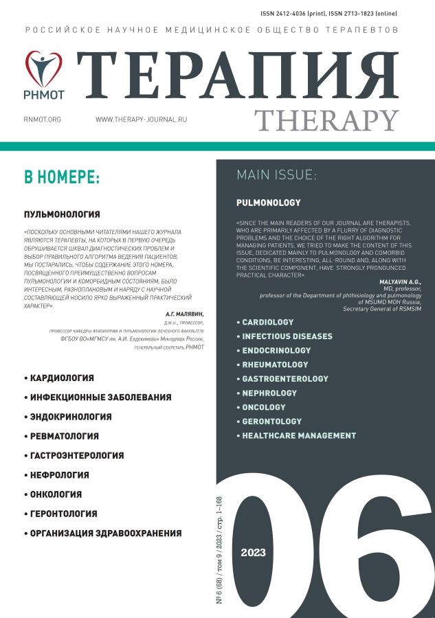
Idiopathic pulmonary hypertension (autopsy observation)
- Autores: Vasyukova O.A.1, Tsareva N.A.2,3, Chernyaev A.L.1,2,4,5, Mikhaleva L.M.1, Samsonova M.V.2,4,6
-
Afiliações:
- B.V. Petrovsky Russian Research Center of Surgery
- Research Institute of Pulmonology of FMBA of Russia
- I.M. Sechenov First Moscow State Medical University of the Ministry of Healthcare of Russia (Sechenov university)
- Central Research Institute of Tuberculosis
- N.I. Pirogov Russian National Research Medical University of the Ministry of Healthcare of Russia
- A.S. Loginov Moscow Clinical Research Center of the Department of Healthcare of Moscow
- Edição: Volume 9, Nº 6 (2023)
- Páginas: 82-88
- Seção: CLINICAL CASES
- URL: https://journals.eco-vector.com/2412-4036/article/view/624547
- DOI: https://doi.org/10.18565/therapy.2023.6.82–88
- ID: 624547
Citar
Texto integral
Resumo
The article presents a description of idiopathic pulmonary arterial hypertension (IPAH) autopsy observation. The diagnosis was made in a 51-year-old patient four years ago and based on the results of an invasive measurement of mean pulmonary artery pressure during right heart catheterization, the index of which was 36 mmHg. During dynamic observation, according to echocardiography, the values of systolic pressure in the pulmonary artery ranged from 66 to 107 mm Hg. Observed patient received treatment with macitentan and spironolactone for 4 years, died with symptoms of pulmonary heart disease. At autopsy, idiopathic plexiform arteriopathy was diagnosed in combination with Heth–Edwards class 4–5 thrombotic arteriopathy with right ventricular failure development.
Texto integral
Sobre autores
Olesya Vasyukova
B.V. Petrovsky Russian Research Center of Surgery
Autor responsável pela correspondência
Email: o.vas.93@gmail.com
ORCID ID: 0000-0001-6068-7009
junior researcher at the Laboratory of clinical morphology, Scientific Research Institute of Human Morphology named after academician A.P. Avtsyn
Rússia, 117418, Moscow, 3 Tsyurupy Str.Natalya Tsareva
Research Institute of Pulmonology of FMBA of Russia; I.M. Sechenov First Moscow State Medical University of the Ministry of Healthcare of Russia (Sechenov university)
Email: tsareva_n_a@staff.sechenov.ru
ORCID ID: 0000-0001-9357-4924
phd in medical sciences, head of the laboratory of intensive care and respiratory failure, associate professor of the department of pulmonology
Rússia, 115682 Moscow, 28 Orekhovy Boulevard; MoscowAndrey Chernyaev
B.V. Petrovsky Russian Research Center of Surgery; Research Institute of Pulmonology of FMBA of Russia; Central Research Institute of Tuberculosis; N.I. Pirogov Russian National Research Medical University of the Ministry of Healthcare of Russia
Email: cheral12@gmail.com
ORCID ID: 0000-0003-0973-9250
md, professor, head of the department of fundamental pulmonology, leading researcher at the Laboratory of clinical morphology, professor of the department of pathological anatomy and clinical pathological anatomical anatomy of the faculty of general medicine, pathologist, Scientific Research Institute of Human Morphology named after academician A.P. Avtsyn
Rússia, 115682, Moscow, 28 Orekhovy Boulevard; Moscow; Moscow; MoscowLyudmila Mikhaleva
B.V. Petrovsky Russian Research Center of Surgery
Email: mikhalevaLM@yandex.ru
ORCID ID: 0000-0003-2052-914X
md, professor, corresponding member of the Russian Academy of Sciences, director, head of the laboratory of clinical morphology, Scientific Research Institute of Human Morphology named after academician A.P. Avtsyn
Rússia, 117418, Moscow, 3 Tsyurupy Str.Maria Samsonova
Research Institute of Pulmonology of FMBA of Russia; Central Research Institute of Tuberculosis; A.S. Loginov Moscow Clinical Research Center of the Department of Healthcare of Moscow
Email: sama-ry@mail.ru
ORCID ID: 0000-0001-8170-1260
md, head of the laboratory of pathological anatomy, senior researcher
Rússia, 115682, Moscow, 28 Orekhovy Boulevard; Moscow; MoscowBibliografia
- Черняев А.Л., Самсонова М.В. Легочная гипертензия (под ред. С.Н. Авдеева). Руководство для врачей. Под ред. члена-корреспондента РАН С.Н. Авдеева. 2-е издание, переработанное и дополненное. М.: ГЭОТАР-Медиа. 2019; 608 с. [Cherniaev A.L., Samsonova M.V. Pulmonary hypertension. Guide for doctors. Ed. by corresponding member of RAS Avdeev S.N. 2nd edition, revised and enlarged. Moscow: GEOTAR-Media. 2019; 608 pp. (In Russ.)]. ISBN: 978-5-9704-5000-0.
- Hoper M.M., Bogaard H.J., Condliffe R. et al. Difinitions and diagnosis of pulmonary hypertension. Definitions and diagnosis of pulmonary hypertension. J Am Coll Cardiol. 2013; 62(25 Suppl): D42–50. https://dx.doi.org/10.1016/j.jacc.2013.10.032.
- Galie N., Hoeper M.M., Humbert M. et al. Guidelines for the diagnosis and treatment of pulmonary hypertension. Eur Respir J. 2009; 34(6): 1219–63. https://dx.doi.org/10.1183/09031936.00139009.
- Heath D., Edwards J.E. The pathology of hypertensive pulmonary vascular disease; a description of six grades of structural changes in the pulmonary arteries with special reference to congenital cardiac septal defects. Circulation. 1958; 18(4 Part 1): 533–47.
- https://dx.doi.org/10.1161/01.cir.18.4.533.
- Katzenstein A. A-L. Katzenstein and Askin’s surgical pathology of non-neoplastic lung disease. Vol.13 of series «Major problems in Pathology». Saunders; 3 rd ed. Philadelphia, Toronto. 1997; 332–60.
- Peacock A.J., Murphy N.F., McMurray J.J. et al. An epidemiological study of pulmonary arterial hypertension. Eur Respir J. 2007; 30(1): 104–9. https://dx.doi.org/10.1183/09031936.00092306.
- Humbert M., Morrell N.W., Archer S.L. et al. Cellular and molecular pathobiology of pulmonary arterial hypertension. J Am Coll Cardiol. 2004; 43(12 Suppl S): 13S–24S. https://dx.doi.org/10.1016/j.jacc.2004.02.029.
- Jang S.Y., Kim E.K., Huh J. et al. A retrospective population-based survival study of idiopathic pulmonary arterial hypertension in Korea. J Korean Med Sci. 2022; 37(10): e80. https://dx.doi.org/10.3346/jkms.2022.37.e80.
- Hoeper M.M., Boucly A., Sitbon O. Age, risk and outcomes in idiopathic pulmonary arterial hypertension. Eur Respir J. 2018; 51(5): 1800629. https://dx.doi.org/10.1183/13993003.00629-2018.
- Dong L., He J.G., Shan G.L. et al. [Clinical analysis of 150 patients with idiopathic pulmonary arterial hypertension. Zhonghua Xin Xue Guan Bing Za Zhi. 2012; 40(8): 657–61 (In Chinese)].
- Jamison B.M., Michel R.P. Different distribution of plexiform lesions in primary and secondary pulmonary hypertension. Hum Pathol. 1995; 26(9): 987–93. https://dx.doi.org/10.1016/0046-8177(95)90088-8.
- Jonigk D., Golpon H., Bockmeyer C.L. et al. Plexiform lesions in pulmonary arterial hypertension composition, architecture, and microenvironment. Am J Pathol. 2011; 179(1): 167–79. https://dx.doi.org/10.1016/j.ajpath.2011.03.040.
- Wagenvoort C.A., Wagenvoort N., Draulans-Noe Y. Reversibility of plexogenic pulmonary arteriopathy following banding of the pulmonary artery. J Thorac Cardiovasc Surg. 1984; 87(6): 876–86.
- Galambos C., Sims-Lucas S., Abman S.H., Cool C.D. Intrapulmonary bronchopulmonary anastomoses and plexiform lesions in idiopathic pulmonary arterial hypertension. Am J Respir Crit Care Med. 2016; 193(5): 574–76.
- https://dx.doi.org/10.1164/rccm.201507-1508LE.
- Wagenvoort C.A., Wagenvoort N. Pathology of pulmonary hypertension. John Wiley and sons: New York. 1977; 345 pp.
- Kim N.H., Delcroix M., Jais X. et al. Chronic thromboembolic pulmonary hypertension. Eur Respir J. 2019; 53(1): 1801915.
- https://dx.doi.org/10.1183/13993003.01915-2018.
- Pietra G.G., Edwards W.D., Kay J.M. et al. Histopathology of primary pulmonary hypertension. A qualitative and quantitative study of pulmonary blood vessels from 58 patients in the National Heart, Lung, and Blood Institute, Primary Pulmonary Hypertension Registry. Circulation. 1989; 80(5): 1198–206. https://dx.doi.org/10.1161/01.cir.80.5.1198.
- Wagenvoort C.A., Mulder P.G. Thrombotic lesions in primary plexogenic arteriopathy. Similar pathogenesis or complication? Chest. 1993; 103(3): 844–49. https://dx.doi.org/10.1378/chest.103.3.844.
- Чазова И.Е., Жданов В.С., Веселова С.П., Мареев В.Ю. Патология первичной легочной гипертензии. Архив патологии. 1993; 55(3): 52–55. [Chazova I.E., Zhdanov V.S., Veselova S.P., Mareev V.Yu. Pathology of primary pulmonary hypertension. Arkhiv patologii = Archive of Pathology. 1993; 55(3): 52–55 (In Russ.)].
Arquivos suplementares


