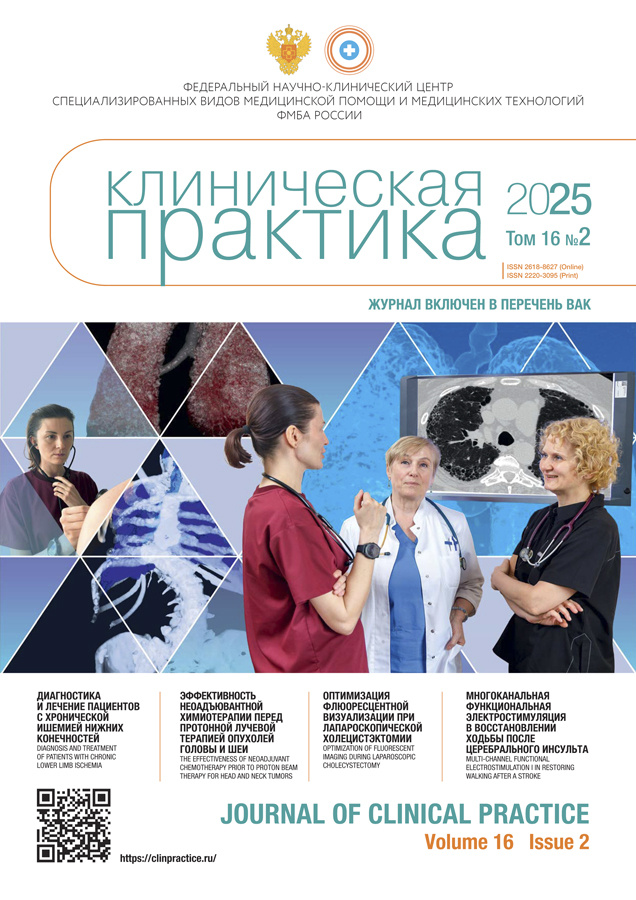
Journal of Clinical Practice
Quarterly peer-review medical journal.
Editor-in-chief
- professor Alexandex I. Troitsky, MD, Dr. Sci. (Medicine)
ORCID: 0000-0003-2143-8696; Scopus Author ID: 57202735847
Main handling editor
- Vladimir P. Baklaushev, MD, Dr. Sci. (Medicine)
ORCID: 0000-0003-1039-4245; Researcher ID: H-2426-2013
Publisher
- Eco-Vector Publishing group
Founder
- Federal Research Clinical Center FMBA of Russia
WEB: https://fnkc-fmba.ru
About
The main idea of our journal is to provide description and analysis of clinical cases with severe, rare and difficult for diagnoses diseases, occurred in the clinics of Federal Medical-Biological Agency of Russia. Such clinical analysis is aimed to develop “clinical” type of thinking, always have been the characteristic feature of Russian/USSR medical school. The journal purpose is also to improve scientific discussions and cooperation between physicians of different specialties.
Revival of historical traditions in our journal is the one of the components of continuing education, which is especially important in “closed” territories, where doctors can`t regularly participate in clinical conferences. An important aspect is to provide a printed tribune for any doctor who has an interesting clinical observation and wish to share his experience with colleagues. That is why we named our journal "Clinical Practice" and address it, first of all, those skilled in applied medicine. Of course, we also publish the results of original researches, clinical guidelines, current reviews and medical news. The journal is multidisciplinary and we hope that it will be interesting to doctors of different specialties. The journal is published by means of the Federal Research and Clinical Center of FMBA of Russia. Placement of all materials, except for advertising, are free of charge to authors.
Types of accepted articles
- reviews;
- systematic reviews and meta-analysis;
- original study articles;
- case reports and series of cases;
- letters to the editor;
- hystorical articles
The joutnal accept manuscripts in English and in Russian.
Publication, distribution and indexation
- Russian and English full-text articles;
- issues publish quarterly, 4 times per year;
- no obligatory APC, Platinum Open Access
- articles distribute under the Creative Commons Attribution-NonCommercial-NoDerivates 4.0 International License (CC BY-NC-ND 4.0).
Indexation
- Russian Science Citation Index (elibrary.ru)
- DOAJ
- CrossRef
- Dimensions
- Google Scholar
- Ulrich's Periodicals Directory
- CyberLeninka
Current Issue
Vol 16, No 2 (2025)
- Year: 2025
- Published: 01.08.2025
- Articles: 13
- URL: https://clinpractice.ru/clinpractice/issue/view/10214
Original Study Articles
Diagnostics and treatment of patients with chronic limb ischemia (single-center experience)
Abstract
BACKGROUND: The success of treating the patients with chronic ischemia of the lower limbs consists of a number of components, such as the full-scale diagnostics; the re-vascularisation volume, the therapeutic means (including the use of modern developments in angiogenesis); the engagement of specialists from the adjacent fields. AIM: the improvement of treatment results in patients with chronic ischemia in the lower limbs and developing an optimal treatment and diagnostic algorithm for this group of patients. METHODS: The analysis included the treatment results of 218 patients with chronic ischemia of the lower limbs (136 males, 82 females; the mean age was 67±6 years), of which 144 patients were operated, 74 were treated conservatively. Diagnostics and examination methods: ultrasound angioscanning, single-photon emission computed tomography combined with three-phase scintigraphy and computed tomography, consulting by a neurologist and a cardiologist, electroneuromyography by prescription from the neurologist and additional examinations by prescription from the cardiologist. The follow-up was implemented as the control out-patient examinations or upon the repeated hospitalization. The follow-up period was 6 months. RESULTS: The distribution of patients by the degree of ischemia as per the classification by А.V. Pokrovsky was the following: IIА — 57 (26.1%), IIB — 31 (14.2%), III — 42 (19.2%), IV — 88 (40.5%) patients. The number of open-access surgeries conducted was 56 (25.7%), with 64 endovascular (29.4%) and 24 hybrid ones (11.0%); conservative therapy was administered to 40 patients (18.3%), conservative therapy accompanied by additional administrations of plasma-free auto-platelet lysate — 34 (15.6%). In the group of operated patients, there were no significant differences depending on the method of surgical treatment at the early post-surgery period and after 6 months of follow-up (p >0.05). Among the patients receiving conservative therapy, the best results at the follow-up of 6 months were reported in patients, in which the standard therapy was accompanied by stimulation of angiogenesis with auto-platelet factors (p <0.05). The presence of ischemic neuropathy was investigated in 98 patients. Neuropathy was detected in 69 cases using the method of electroneuromyography. After prescribing the neurotropic therapy, resolving of neuropathic pain was reported in 52 (75.4%) patients. CONCLUSION: The multi-disciplinary approach developed by us for the diagnostics and treatment of patients with chronic ischemia of the lower limbs, allows for improving the treatment results, while the extended spectrum of diagnostic methods allows for evaluating the risk factors, for determining the optimal treatment tactics and for objectively evaluating the dynamic changes of the patient status.
 7-14
7-14


The effectiveness of neoadjuvant chemotherapy prior to proton beam therapy for head and neck tumors
Abstract
BACKGROUND: Malignant tumors of the head and neck are a significant problem in modern oncology, as they occupy an important place in the structure of morbidity and mortality of the population. According to the Ministry of Health of the Russian Federation, 674,587 new cases of malignant neoplasms were registered in 2023, of which 25,038 cases were tumors of the head and neck. AIM: of the study was to evaluate the effect of induction drug therapy on the treatment outcomes of patients with locally advanced tumors of the head and neck who received radiation treatment using proton therapy, IMPT technique (intensity modulated proton therapy). METHODS: The retrospective study included an analysis of the medical records of 103 patients with head and neck tumors, who were divided into two groups: patients who received induction chemotherapy followed by proton chemoradiotherapy (n=50), and patients who did not receive induction antitumor treatment before starting proton chemoradiotherapy (n=53). T-tests for independent samples were used to assess differences between patient groups. The statistical significance of the differences was considered at a level of p <0.05. RESULTS: The median follow-up was 13.4 months (IQR: 11.6–21.6 months). The average follow-up time was 15.7±7.8 months. In the group of monitored patients, none interrupted planned treatment, and therapy was completed on time. In the induction chemotherapy followed by proton chemoradiation therapy group, the average OS was 27.65 months (95% CI: 24.46–30.85), while for the proton chemoradiation therapy groups it was 27.27 months (95% CI: 22.15–31.72), which was a statistically insignificant difference (Chi-squared 0.776, p=0.378). The median OS for both study groups was not reached. The progression-free survival assessment showed that the average time to progression in the induction chemotherapy followed by proton chemoradiation therapy group was 23.1 months (95% CI: 19.6–26.6), versus 21.2 months (95% CI: 16.7–25.7) in the proton chemoradiation therapy group. The incidence of grade 1 leukopenia was 30% in the induction chemotherapy followed by proton chemoradiation therapy group versus 20.8% in the proton chemoradiation therapy group, the incidence of grade 3 disorders was 26% in the induction chemotherapy followed by proton chemoradiation therapy group and 11.3% in the proton chemoradiation therapy group, and grade 3 complications were noted only in the induction chemotherapy followed by proton chemoradiation therapy group (12%). These differences are statistically significant (p <0.01). CONCLUSION: This study demonstrated that induction chemotherapy does not improve overall survival and progression-free survival in patients with locally advanced squamous cell carcinoma of the head and neck receiving proton chemoradiotherapy.
 15-22
15-22


Automated morphometry of the prostate gland by the results of magnetic resonance imaging
Abstract
BACKGROUND: Within the framework of the experiment on using the innovative technologies in the field of computer vision for analyzing the medical images and on further usage of these technologies in the healthcare system of the City of Moscow, the research was carried out using the equipment based on the artificial intelligence (AI-service) for the purpose of automatization of the morphometry of the prostate gland using the magnetic resonance imaging (MRI), for the issue is topical due to the high incidence of urological diseases among men. Unlike the 11 previous systems, oriented at the retrospective analysis, this solution helps the radiologists in shortening the time of describing the examination results and in increasing their accuracy. AIM: to evaluate the quality and the validity of automatic morphometry of the prostate gland by the MRI results using the technologies of artificial intelligence in the settings of practical healthcare. METHODS: A prospective diagnostic research in accordance with the methodology of reporting results of scientific research involving the STARD 2015 diagnostic tests was conducted during the period from April until October of 2024. A total of 560 MRI results were used and compared to the data from the morphometric AI-service. RESULTS: An evaluation of the accuracy of using the AI-service for the morphometry of the prostate gland was carried out. A total of 7 clinical monitoring procedures were conducted using 560 MRI datasets with the complete conformity reported in 71.6%. The rate of false-negative cases was 3.9%, technical defects were found in 3.8% of the cases. The integral clinical evaluation has achieved the range of 88.0–97.0%, confirming the high diagnostic quality. The predominant errors were the ones related to the contouring of the gland (52%) and incorrect measurements (13%), often related to the prolapsing of the prostate gland apex. CONCLUSION: The automatization of routine measurements greatly contributes to the standardizing the processes of describing the results obtained by radio-diagnostic methods. This aspect is of special importance from the point of view of providing the continuity of medical aid in case of patients presenting to various medical organizations. The artificial intelligence technologies for the automatization of the prostate gland measurements have demonstrated high clinical value in 92.0%, which indicates their accuracy and quality. These data can be used for developing new MRI-based automated morphometry products.
 23-33
23-33


Optimizing the fluorescent visualization with Indocyanine green during laparoscopic cholecystectomy
Abstract
BACKGROUND: The prevention of damaging the bile ducts during surgical interventions in patients with calculous cholecystitis remains a topical problem in modern abdominal surgery. The incidence of damaging the bile ducts reaches 0.4–2%, while in cases of complicated forms — up to 5.2%. AIM: determining the optimal dosage and timing of administering the Indocyanine green (ICG) for increasing the efficiency of fluorescent cholangiography during the course of laparoscopic cholecystectomy in cases of calculous cholecystitis. The top-priority task of the research is minimizing the risks of injuring the bile ducts by means of clear intraoperative visualization of the extrahepatic bile ducts. METHODS: Prospective non-randomized research was conducted within the premises of the University Clinical Center named after V.V. Vinogradov (affiliated branch) of the RUDN University during the period from March 2024 until April 2025. The research included 276 patients undergoing the laparoscopic cholecystectomy with using the ICG-cholangiography. The dosages of Indocyanine green used (1.25 mg; 2.5 mg; 5 mg; 10 mg) were administered in various time periods before starting the surgery (from 40 minutes up to 6 hours), as well as intraoperatively. The evaluation included the degree of fluorescence, the time from the moment of administering the Indocyanine green until the optimal fluorescence of the bile ducts and of the liver required for the safe conduction of laparoscopic cholecystectomy, as well as the possibility to correct the hyper- and hypofluorescence by changing the equipment settings. RESULTS: Optimal visualization of the extrahepatic bile ducts was observed at a dosage of 5 mg of ICG 3–5 hours after the administration, for the 2.5 mg dosage — 2–3 hours, while for the 1.25 mg dosage, the time was 40–120 minutes. Intraoperative administration of 1.25 mg provided a rapid visualization, but caused hyperfluorescence, complicating the determination of the topography of bile ducts, which was corrected by equipment settings. CONCLUSION: Fluorescent cholangiography with using the Indocyanine green is a safe and effective method of visualizing the extrahepatic bile ducts during laparoscopic cholecystectomy. The most optimal dosages of Indocyanine green are the following: 1.25 mg — 40–120 minutes, 2.5 mg — 2–3 hours and 5 mg — 3–5 hours before the intervention. The 1.25 mg dosage can be administered intraoperatively with further correction of equipment settings at the menu of the video-system (by lowering the enhancement and the intensity parameters) for decreasing the hyperfluorescence effect.
 34-40
34-40


The evaluation of efficiency of the impact of vitamin D and remineralizing toothpaste on the structure of the dental enamel in the individuals with homozygous polymorphism in the gene, encoding the intracellular vitamin D receptor (VDR)
Abstract
BACKGROUND: Only a few literature sources data show the relation of the VDR gene polymorphism and the susceptibility to developing dental caries. Within this context, investigating the structure of the dental enamel and the changes of its resistance under the effects of vitamin D and remineralizing therapy among the persons with the homozygous polymorphism of the VDR gene (А/A) is topical. AIM: to investigate the effects of vitamin D and the toothpaste with remineralizing contents on the structure of the enamel surface of the impacted teeth extracted from the individuals with homozygous polymorphism of the VDR gene. METHODS: In 2023–2025, within the premises of the Dentistry Department of the Federal State Budgetary Institution of Continuing Professional Education “Central State Medical Academy”, a total of 200 students aged 22–25 years were screened with undergoing a genetic testing to reveal the polymorphism of the VDR gene. Out of the 36 assessed subjects, 18 individuals were detected with the homozygous А/А allele that are currently undergoing orthodontic therapy and requiring an extraction of the impacted molars. A total 24 of extracted teeth were tested with submerging them into the artificial saliva with an addition of various media. The dental samples were distributed into four groups: only artificial saliva (I, control); 1000 IU of cholecalciferol per 100 ml (II); processing with remineralizing toothpaste (III); vitamin D and remineralizing toothpaste (IV). The evaluation of the structure of the dental enamel was carried out using the method of confocal profilometry with measuring the Ra and Rp roughness parameters. RESULTS: In group II with the presence of cholecalciferol, changes were revealed in the roughness parameters (Ra, Rр) of dental enamel surface, in group III (processing with remineralizing toothpaste) the Ra and Rр parameters had similar digital values. As for the samples from the group IV, comparing to the group I, smoothness was revealed in the dental enamel surface, which is confirmed by the Rа and Rр (p >0.001) parameters. This effect can be explained by the synergic action of the cholecalciferol and the remineralizing components of the toothpaste on the structure of the enamel. CONCLUSION: In the individuals with homozygous polymorphism (А/А) of the VDR gene, significant changes were revealed in the parameters of dental enamel roughness (Ra and Rр) after the combined use of cholecalciferol and remineralizing toothpaste, which is related to the smoothening of surface due to the formation of the homogeneous layer consisting of the microRepair microcrystals.
 41-52
41-52


Reviews
Combined physical activity in the prevention of cardiovascular diseases: from physiological to molecular adaptation mechanisms
Abstract
Physical activity is recognized as the most important non-medicinal tool for the prevention of cardiovascular diseases, however, the highest efficiency is demonstrated by the programs combining various types of physical load. The present review summarizes the current scientific data on the effects of combined physical activity, including the aerobic exercises of moderate and high intensity and the muscle strengthening exercises, as well as the ones affecting the system of cardiometabolic regulation. A disclosure is provided for the multi-level adaptation mechanisms, encompassing the physiological, vegetative, hormonal, molecular and epigenetic levels. It was justified that combined training modalities possess the synergetic effects, facilitating the decrease of blood pressure, the increase in the cardiac rhythm variability, the improvement of insulin sensitivity, the decrease of chronic inflammation and the activation of cardioprotective transcription programs. A detailed description was provided for the key molecular pathways participating in the adaptational response (AMP-activated protein kinase, mTOR, PGC-1α, autophagia and the unfolded protein response), as well as the role of exerkines — the signaling molecules produced in response to physical load. Special attention was paid to the epigenetic modifications, including the methylation of DNA, the regulation of microRNA and the telomere stability, as the mechanisms of long-term protection from the cardiovascular diseases. The data provided emphasizes the necessity of introducing the combined modalities of physical activity into the programs of individual and populational prevention. Besides, further research is required on the optimal combination of intensity, extent and patterns of physical load for various population categories for the purpose of maximizing the protective adaptation of the cardiovascular system.
 53-68
53-68


Multi-channel functional electrostimulation: the method of restoring the walking function in patients with a past history of acute cerebrovascular event
Abstract
Multi-channel functional electrostimulation (MFES) represents a promising method for the rehabilitation of post-stroke patients, aimed at restoring the walking function in various periods after an acute cerebrovascular event. The review systematizes the modern concepts of using the MFES in patients with the consequences of cerebral stroke, analyzing the technical parameters of stimulation, the methodical approaches to conducting the procedures and the clinical efficiency of the method. The analysis of literature data demonstrates significant variability of MFES protocols: the stimulation frequency varies from 20 to 100 Hz, the duration of the procedure ranges from 15 to 60 minutes, the treatment course can last from 3 to 30 weeks. The main targets of stimulation are the four groups of muscles in the lower limbs — the anterior tibial muscle, the plantar flexors, the quadriceps muscle of thigh and the group of muscles on the posterior surface of thigh. The synchronization of stimulation with the walking cycle is conducted predominantly by means of contact sensors, accelerometers and electromyographic signals; modern developments include the inertial systems and the machine learning algorithms. The review presents a combined analysis of the technical aspects of MFES from the point of view of staging of motor learning and individualization of the stimulation parameters. Special attention was paid to the integration of MFES with the robotic devices, including the exoskeletons, which represents a new trend in rehabilitation. Along with the absence of the unified criteria for choosing the stimulation parameters, it is worth noting that there is a necessity of differentiated approach depending on the type of motor disorders, on the duration of the disease and on the cognitive capabilities of the patient. The analysis presented justifies the necessity of developing personalized MFES protocols and arranging a large-scale research for optimizing the stimulation parameters in the rehabilitation of post-stroke patients.
 69-81
69-81


Suprascapular neuropathy combined with massive rotator cuff tears: clinical signs, diagnostics, treatment
Abstract
Suprascapular neuropathy is a type of disease, which up until recently was considered quite rare and observed mainly among the athletes. On the contrary, this problem is observed quite often, especially as a professional disease among the individuals involved in heavy manual labour. The syndrome of suprascapular nerve compression is a complex problem, combining a multitude of reasons and resulting in the atrophy of the supraspinal and infraspinous muscles. The compression of the suprascapular nerve is developing due to its complex anatomy, the presence of additional bony and other types of structures in the area of the scapular notch, as well as due to the traumatic lesions of the rotator cuff and of the scapular spine. There is an opinion that the contraction of the damaged supraspinal and infraspinous muscles may cause contusion- related changes in the suprascapular nerve, which may persist after the reconstruction of the rotator cuff. The provided research summarizes the data available on the suprascapular neuropathy, especially combined with massive rotator cuff tears, as well as on the reasons of its development, the clinical manifestations, the diagnostics and the comparative results for various treatment methods. An analysis was conducted of the main research results obtained using the surgical treatment for suprascapular neuropathy, in particular, the arthroscopic decompression combined with the treatment of rotator cuff abnormalities. The analysis of literature data has shown that, in case of the presence of space-occupying masses or bone deformities in the area of the scapular notch, surgical correction shows significant positive results. In case of damaged rotator cuff, the combination of arthroscopic release of the suprascapular nerve with its reconstruction provides good clinical results, promoting to the decrease of the pain syndrome during the postoperative period, however, no significant differences were reported when restoring the rotator cuff both with the release procedure and without it. Most part of the patients with chronic pain syndrome and degenerative changes can be successfully treated conservatively. Despite the fact that the relation of rotator cuff tears and suprascapular neuropathy is undoubtful, many researchers describe the absence of statistically significant difference in the clinical results of reconstructing the rotator cuff together with arranging the procedure of arthroscopic release and without it. Thus, probably, the indications to arthroscopic release of the suprascapular nerve should be clearly limited to cases of its neuropathy based on the data obtained during the research including larger samples of patients.
 82-89
82-89


Pneumonitis as a complication of Immunotherapy of oncological diseases: difficulties in diagnosis and treatment
Abstract
Pneumonitis is one of the life-threatening complications of immunotherapy of oncological diseases. Despite the low occurrence rate, pneumonitis significantly affects not only the quality of life in the patients, but also the mortality, forcing to change the treatment scheme of the main disease. Therapy with immune checkpoint blockers is a relatively new, but well established type of oncology therapy. It is expected that, with the extension of the list of indications to immunotherapy, some growth would be observed in the number of complications, due to which it becomes necessary to inform the physicians of various (not only oncological) specialties about it. It is important for them to maintain high index of suspicion to be able to detect this life-threatening complication at the early stages and to prescribe adequate treatment. The currently known research works by national and foreign authors on the detection and treatment of pneumonitis in their majority have a strictly specialized type due to studying a specific immunotherapy medicine or a specific location of the tumor. In the present article, we have summarized and systematized the data from various sources, emphasizing on the diagnostics and therapy of this dangerous complication.
 90-96
90-96


Case reports
Difficulties in the diagnostics of transthyretin amyloidosis with polyneuropathy: a clinical case description
Abstract
BACKGROUND: In clinical practice, diagnosing of systemic transthyretin amyloidosis (ATTR-amyloidosis) with the impairment of the nervous and cardiovascular systems became possible due to the accessibility of genetic diagnostics and due to the growth of knowledge on this disease. CLINICAL CASE DESCRIPTION: A clinical case is presented of transthyretin amyloidosis, manifesting as polyneuropathy with significant neuropathic pain syndrome, with the development of severe vegetative insufficiency and with the impairment of the cardiovascular system in a 63 years old male. This clinical pattern, developing for 3 years, was associated with multiple encounters of the patient to various specialists — rheumatology physicians, neurosurgery specialists, endocrinologists, neurologists, internists, including surgical interventions in the spinal cord, carotid artery and cardiac vessels. The disease has lead to the development of severe cachexia and incapacitation of the patient. CONCLUSION: Taking into consideration that ATTR-amyloidosis manifests with the clinical signs of lesions in various organs and systems, and this is interpreted by the specialists within the framework of their specific field apart from the general etiology of the disease. The patient gets prescribed with multiple examinations and symptomatic medications, which causes a delay in the diagnostics and in setting the correct diagnosis. This clinical case describes the classical form of transthyretin amyloidosis, which, upon timely diagnostics, has its pathogenetic therapy.
 97-103


The patient’s way to diagnosis: a clinical case of late onset Pompe disease in an adult
Abstract
BACKGROUND: Late onset Pompe disease is a rare genetic disease from the group of the accumulation diseases, the main manifestation of which is the progressive myopathic syndrome. The difficulties of diagnostics are mainly related to the low awareness of the specialized physicians (neurologists, orthopaedists, rheumatologists, pulmonologists, pediatricians etc.) on the specific features of this disease, as well as to the low level of nosological specificity of the myopathic syndrome in general. Special importance of early diagnostics is due to the existence of pathogenetic therapy. Late diagnostics and delayed initiation of therapy lead to lower survival rate and higher rate of incapacitation among the patients. CLINICAL CASE DESCRIPTION: The patient aged 62 years, which for many years was under the supervision by neurologists with the diagnosis of osteochondrosis, based on the objective data, actually had a myopathic syndrome that was diagnosed and confirmed using the electroneuromyography. The detected findings included a decrease in the activity of the alpha-glucosidase enzyme, while the genetic examination that followed, allowed for detecting the presence of a mutation in the GAA gene. The dynamic changes of the disease were tracked with a background of taking pathogenetic therapy for 4 years. CONCLUSION: This clinical case demonstrates the many years of the patient’s way to being diagnosed with a rare genetic disease and, respectively, to the later initiation of therapy. The efficiency of treating the accumulation diseases directly depends on the extent of the pathological changes in the target organs, accumulated to the moment of diagnostics, which means — the earlier, the more effective.
 104-111


The combined approach in the treatment of a patient with glaucoma and endothelial-epithelial corneal dystrophy: a clinical case description
Abstract
BACKGROUND: Penetrating keratoplasty provides a possibility of restoring vision in patients with various corneal diseases, however, just like any surgical intervention, the operation is associated with certain risks and has a number of contraindications. One of the unfavorable prognostic factors in cases of penetrating keratoplasty is uncompensated glaucoma. Penetrating keratoplasty can result in the reactive postoperative hypertension, however, this is not a standard situation. In patients with a history of glaucoma, this complication occurs much more often than in patients without previously diagnosed glaucoma. The increase of intraocular pressure during the postoperative period in patients suffering from glaucoma, can lead to the progression of the disease and to the development of the transplant disease. CLINICAL CASE DESCRIPTION: This article presents a clinical case of a patient with juvenile glaucoma, which underwent several glaucoma surgeries and later he received an Ex-PRESS implanted glaucoma drainage. The drainage implant was in contact with the posterior surface of the cornea, as a result of which, initially local and further total endothelial-epithelial corneal dystrophy has developed with the formation of stromal cloudiness and progressing of pain syndrome. The drainage was removed and subsequently a decision was drawn up on arranging a penetrating corneal keratoplasty, for the critical flicker fusion rate was 30 Hz, which allowed for expecting a sufficiently high vision acuity during the postoperative period. However, despite the maximal hypotensive regimen, the intraocular pressure remained high, due to which, for the purpose of decreasing it before corneal transplantation, a transscleral diode laser cyclophotocoagulation was used. CONCLUSION: The presented clinical case demonstrates the efficiency of transscleral diode laser cyclophotocoagulation in a patient with uncompensated glaucoma in terms of the quality of preparation to penetrating keratoplasty.
 112-118


Implantation of an additional intraocular lens for keratoconus in a pseudophakic eye
Abstract
BACKGROUND: In the accessible literature sources, there is insufficient information on the correction of refraction abnormalities in cases of keratoconus, due to which exploring the modern approaches in the implantation of additional intraocular lenses, including the choice of indications, the surgical technique and the post-operative follow-up, gains major importance for managing the patients with this disease. CLINICAL CASE DESCRIPTION: The patient G., aged 42 years, presented with the complaints of decreased visual acuity in the left eye. Past medical history of progressing decreased visual acuity in both eyes from 2018, the diagnosis set was the following: “Right eye (OD): keratoconus stage I, left eye (OS): keratoconus stage II”. In 2018, the implantation of intrastromal corneal ring segments in both eyes was conducted, in 2019 — refractive lensectomy with the implantation of the AcrySof IQ Toric SN6AT8 intraocular lens (Alcon, USA) in both eyes. The examination results in the OS upon presenting were the following: non-corrected visual acuity 0.05, maximum corrected visual acuity 0.5; autorefractometry: sph +2.25 D; cyl -9.50 D ax 81°; intraocular pressure — 17 mm.Hg. For correcting the refractive error that is preventing from achieving the high visual acuity (far vision), the implantation of additional intraocular lenses was carried out (Sulcofix Toric Care group, India). The results of examining the OS during the first 24 hours after surgery were the following: non-corrected visual acuity of the OS 0.8; autorefractometry: sph -0.25 D; cyl -14.50 D ax 81°; intraocular pressure — 17 mm.Hg. CONCLUSION: The implantation of additional Sulcofix Toric intraocular lenses have demonstrated its efficiency in correcting the refractive error in the pseudophakic eye with keratoconus, however, due to the irregular astigmatism characteristic for keratoconus, the residual defect can still persist.
 119-126


