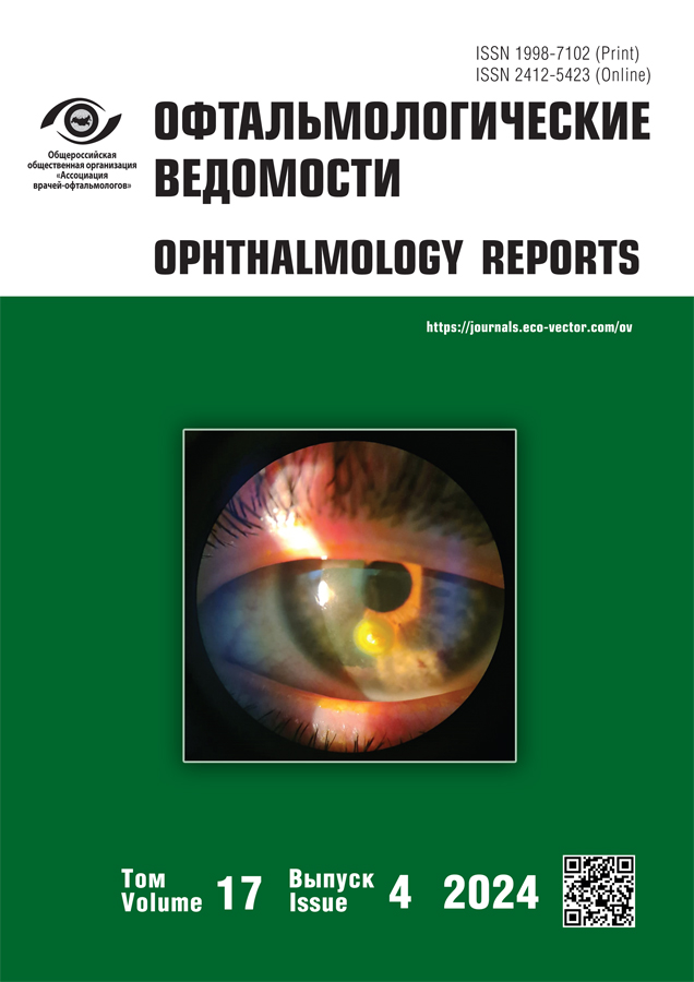
Замена торической интраокулярной линзы на монофокальную у пациента с субэпителиальной фиброплазией после радиальной кератотомии. Клинический случай
- Авторы: Малюгин Б.Э.1,2,3, Калинникова С.Ю.1, Ткаченко И.С.1, Халецкая А.А.1, Меловацкий П.Д.2
-
Учреждения:
- Национальный медицинский исследовательский центр «Межотраслевой научно-технический комплекс «Микрохирургия глаза» им. акад. С.Н. Фёдорова»
- Российский университет медицины
- Глазной Институт Джулиус Стайн
- Выпуск: Том 17, № 4 (2024)
- Страницы: 87-98
- Раздел: Клинические случаи
- Статья получена: 23.04.2024
- Статья одобрена: 28.10.2024
- Статья опубликована: 30.12.2024
- URL: https://journals.eco-vector.com/ov/article/view/630721
- DOI: https://doi.org/10.17816/OV630721
- ID: 630721
Цитировать
Аннотация
Представленный клинический случай демонстрирует замену торической интраокулярной линзы на монофокальную вследствие неверной оценки топографии роговицы у пациента с радиальными рубцами роговицы после кератотомии. Торическая интраокулярная линза была рассчитана по исходным параметрам роговицы, когда её центральная оптическая зона была прозрачна, а на средней периферии в районе 4–5 мм от центральной оптической зоны в верхненаружном квадранте имелась субэпителиальная фиброплазия. Послеоперационный рефракционный результат был не оптимальным, отмечался остаточный астигматизм, который было решено нивелировать скарификацией эпителия в зоне фиброплазии. Это привело к значительному изменению кератометрии и практически полному восстановлению сферичной формы роговицы. Пациенту выполнена замена ИОЛ, что привело к повышению зрительных функций.
Полный текст
Об авторах
Борис Эдуардович Малюгин
Национальный медицинский исследовательский центр «Межотраслевой научно-технический комплекс «Микрохирургия глаза» им. акад. С.Н. Фёдорова»; Российский университет медицины; Глазной Институт Джулиус Стайн
Автор, ответственный за переписку.
Email: boris.malyugin@gmail.com
ORCID iD: 0000-0001-5666-3493
SPIN-код: 8906-2787
д-р мед. наук, профессор, чл.-корр. РАН, заслуженный деятель науки Российской Федерации, председатель Общества офтальмологов России
Россия, Москва; Москва; Лос-Анжелес, СШАСветлана Юрьевна Калинникова
Национальный медицинский исследовательский центр «Межотраслевой научно-технический комплекс «Микрохирургия глаза» им. акад. С.Н. Фёдорова»
Email: svkalinnikova@gmail.com
ORCID iD: 0000-0002-9109-2400
SPIN-код: 6733-2260
MD
Россия, МоскваИван Сергеевич Ткаченко
Национальный медицинский исследовательский центр «Межотраслевой научно-технический комплекс «Микрохирургия глаза» им. акад. С.Н. Фёдорова»
Email: dr.ivan.tka@gmail.com
ORCID iD: 0000-0003-1756-7911
SPIN-код: 9756-8239
MD
Россия, МоскваАнастасия Андреевна Халецкая
Национальный медицинский исследовательский центр «Межотраслевой научно-технический комплекс «Микрохирургия глаза» им. акад. С.Н. Фёдорова»
Email: khaletskaya261@gmail.com
ORCID iD: 0000-0002-4775-9423
MD
Россия, МоскваПавел Дмитриевич Меловацкий
Российский университет медицины
Email: melovatskiy.pavel@gmail.com
ORCID iD: 0000-0002-3500-2220
MD
Россия, МоскваСписок литературы
- Li A., He Q., Wei L., et al. Comparison of visual acuity between phacoemulsification and extracapsular cataract extraction: a systematic review and meta-analysis // Ann Palliat Med. 2022. Vol. 11, N 2. P. 551–559. doi: 10.21037/apm-21-3633
- Малюгин, Б.Э. Медико-технологическая система хирургической реабилитации пациентов с катарактой на основе ультразвуковой факоэмульсификации с имплантацией интраокулярной линзы: автореф. дис. … д-ра мед. наук. Москва, 2002. 50 с. EDN: ZLCMWR
- Chung J., Bu J.J., Afshari N.A. Advancements in intraocular lens power calculation formulas // Curr Opin Ophthalmol. 2022. Vol. 33, N 1. P. 35–40. doi: 10.1097/ICU.0000000000000822
- Narang R., Agarwal A. Refractive cataract surgery // Curr Opin Ophthalmol. 2024. Vol. 35, N 1. P. 23–27. doi: 10.1097/ICU.0000000000001005
- Taban M., Behrens A., Newcomb R.L., et al. Acute endophthalmitis following cataract surgery: a systematic review of the literature // Arch Ophthalmol. 2005. Vol. 123, N 5. P. 613–620. doi: 10.1001/archopht.123.5.613
- Fu L., Patel B.C. Radial Keratotomy Correction. In: StatPearls [Internet]. Treasure Island (FL): StatPearls Publishing; July 24, 2023.
- Воронин Г.В., Бубнова И.А. Изменения биомеханических свойств роговицы после кераторефракционных вмешательств // Вестник офтальмологии. 2019. Т. 135, № 4. С. 108–112. EDN: XTUZLL doi: 10.17116/oftalma2019135041108
- Moshirfar M., Ostler E.M., Smedley J.G., et al. Age of cataract extraction in post-refractive surgery patients // J Cataract Refract Surg. 2014. Vol. 40, N 5. P. 841–842. doi: 10.1016/j.jcrs.2014.03.001
- Leite de Pinho Tavares R., de Almeida Ferreira G., Ghanem V.C., Ghanem R.C. IOL power calculation after radial keratotomy using the Haigis and Barrett True-K formulas // J Refract Surg. 2020. Vol. 36, N 12. P. 832–837. doi: 10.3928/1081597X-20200930-02
- Curado S.X., Hida W.T., Vilar C.M.C., et al. Intraoperative aberrometry versus preoperative biometry for IOL power selection after radial keratotomy: a prospective study // J Refract Surg. 2019. Vol. 35, N 10. P. 656–661. doi: 10.3928/1081597X-20190913-01
- Stakheev A.A. Intraocular lens calculation for cataract after previous radial keratotomy // Ophthalmic Physiol Opt. 2002. Vol. 22, N 4. P. 289–295. doi: 10.1046/j.1475-1313.2002.00033.x
- Wang L., Spektor T., de Souza R.G., Koch D.D. Evaluation of total keratometry and its accuracy for intraocular lens power calculation in eyes after corneal refractive surgery // J Cataract Refract Surg. 2019. Vol. 45, N 10. P. 1416–1421. doi: 10.1016/j.jcrs.2019.05.020
- Turnbull A.M.J., Crawford G.J., Barrett G.D. Methods for intraocular lens power calculation in cataract surgery after radial keratotomy // Ophthalmology. 2020. Vol. 127, N 1. P. 45–51. doi: 10.1016/j.ophtha.2019.08.019
- Альноелати-Альмасри М.А., Стебнев В.С. Торические интраокулярные линзы: исторический обзор, отбор пациентов, расчёт ИОЛ, хирургическая техника, клинический исход и осложнения // Национальная ассоциация ученых. 2021. № 36. С. 16–28. EDN: JBEJPT
- Colombo-Barboza G.N., Rodrigues P.F., Colombo-Barboza F.D.P., et al. Radial keratotomy: background and how to manage these patients nowadays // BMC Ophthalmol. 2024. Vol. 24, N 1. P. 9. doi: 10.1186/s12886-023-03261-0
- Parmley V., Ng J., Gee B., et al. Penetrating keratoplasty after radial keratotomy. A report of six patients // Ophthalmology. 1995. Vol. 102, N 6. P. 947–950. doi: 10.1016/s0161-6420(95)30929-3
- Mohankumar A., Mohan S. Toric intraocular lenses. In: StatPearls [Internet]. Treasure Island (FL): StatPearls Publishing; July 3, 2023.
- Joshi V.P., Chatterjee S., Basu S. Relationship of density, depth, and surface irregularity of superficial corneal opacification with visual acuity // Curr Eye Res. 2023. Vol. 48, N 6. P. 536–545. doi: 10.1080/02713683.2023.2173786
- Almulhim A., Magliyah M.S., Alfawaz A., et al. Successful surgical management of post-penetrating or deep lamellar keratoplasty Acquired Corneal Sub-Epithelial Hypertrophy (ACSH): A case series // Int J Surg Case Rep. 2020. Vol. 67. P. 191–195. doi: 10.1016/j.ijscr.2020.01.054
- Raber I.M., Eagle R.C. Jr. Peripheral hypertrophic subepithelial corneal degeneration // Cornea. 2022. Vol. 41, N 2. P. 183–191. doi: 10.1097/ICO.0000000000002716
- Труфанов С.В., Рикс И.А., Эзугбая М. Узловая дегенерация Зальцмана // Офтальмология. 2022. Т. 19, № 3. С. 482–488. EDN: RKZRAE doi: 10.18008/1816-5095-2022-3-482-488
- Maust H.A., Raber I.M. Peripheral hypertrophic subepithelial corneal degeneration // Eye Contact Lens. 2003. Vol. 29, N 4. P. 266–269. doi: 10.1097/01.icl.0000087489.61955.82
- Gunzinger J.M., Voulgari N., Petrovic A., et al. Peripheral hypertrophic subepithelial corneal degeneration: clinical aspects related to in vivo confocal microscopy and optical coherence tomography // Int Med Case Rep J. 2019. Vol. 12. P. 237–241. doi: 10.2147/IMCRJ.S208297
- Melles G.R., Binder P.S., Anderson J.A. Variation in healing throughout the depth of long-term, unsutured, corneal wounds in human autopsy specimens and monkeys // Arch Ophthalmol. 1994. Vol. 112, N 1. P. 100–109. doi: 10.1001/archopht.1994.01090130110027
- Lagali N., Germundsson J., Fagerholm P. The role of Bowman’s layer in corneal regeneration after phototherapeutic keratectomy: a prospective study using in vivo confocal microscopy // Invest Ophthalmol Vis Sci. 2009. Vol. 50, N 9. P. 4192–4198. doi: 10.1167/iovs.09-3781
- Lee S.J., Sun H.J., Choi K.S., Park S.H. Intraocular lens exchange with removal of the optic only // J Cataract Refract Surg. 2009. Vol. 35, N 3. P. 514–518. doi: 10.1016/j.jcrs.2008.11.045
- Karamaounas N., Kourkoutas D., Prekates C. Surgical technique for small-incision intraocular lens exchange // J Cataract Refract Surg. 2009. Vol. 35, N 7. P. 1146–1149. doi: 10.1016/j.jcrs.2009.02.036
- Geggel H.S. Simplified technique for acrylic intraocular lens explantation // Ophthalmic Surg Lasers. 2000. Vol. 31, N 6. P. 506–507.
- Kuo Y.W., Hou Y.C. Late intraocular lens exchange in dissatisfied patients with multifocal intraocular lens implantation // Taiwan J Ophthalmol. 2020. Vol. 12, N 1. P. 109–112. doi: 10.4103/tjo.tjo_55_20
- Lee M.H., Webster D.L. Intraocular lens exchange-removing the optic intact // Int J Ophthalmol. 2016. Vol. 9, N 6. P. 925–928. doi: 10.18240/ijo.2016.06.23
- Yu A.K., Ng A.S. Complications and clinical outcomes of intraocular lens exchange in patients with calcified hydrogel lenses // J Cataract Refract Surg. 2002. Vol. 28, N 7. P. 1217–1222. doi: 10.1016/s0886-3350(02)01357-3
- Stewart S.A., McNeely R.N., Chan W.C., Moore J.E. Visual and refractive outcomes following exchange of an opacified multifocal intraocular lens // Clin Ophthalmol. 2022. Vol. 16. P. 1883–1891. doi: 10.2147/OPTH.S3629
Дополнительные файлы


