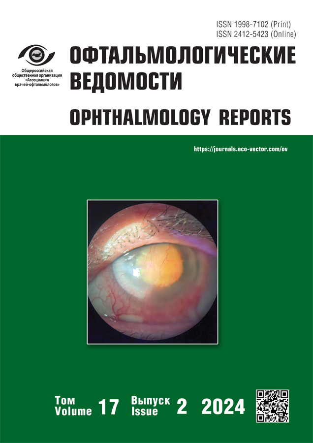
Multifocal character of lesions in gunshot open globe injury type B in experiment
- Authors: Kol'bin A.A.1, Kulikov A.N.1, Zybina N.N.2, Frolova M.Y.2, Troyanovsky R.L.1, Chirskiy V.S.1
-
Affiliations:
- Kirov Military Medical Academy
- Nikiforov All-Russian Center Emergency and Radiation Medicine
- Issue: Vol 17, No 2 (2024)
- Pages: 67-80
- Section: Experimental trials
- Submitted: 16.02.2024
- Accepted: 15.05.2024
- Published: 24.06.2024
- URL: https://journals.eco-vector.com/ov/article/view/627078
- DOI: https://doi.org/10.17816/OV627078
- ID: 627078
Cite item
Abstract
BACKGROUND: An increase was noted in the number of gunshot eyeball injuries, which are accompanied by low functional outcomes. Reproduction and experimental study of this type of eye injury would help improving the functional and cosmetic treatment results in patients.
AIM: The aim of the study is to investigate gunshot open globe injury type B (penetrating wound without intraocular foreign body) on a standardized experimental model.
MATERIALS AND METHODS: A complete investigation of the standardized model of gunshot open globe injury type B (penetrating wound without intraocular foreign body) simulated on the ballistic test facility was carried out. The experiment was accomplished at the ophthalmology chair on 36 rabbits (71 eyes). The injury was inflicted in the projection of the ciliary body — zone II (Birmingham Eye Trauma Terminology). The examination in the control period included ophthalmologic (ophthalmoscopy, full field electroretinography, optical coherence tomography), biochemical (testing of vitreous fibronectin level), histological and radiological (magnetic resonance imaging, ultrasound examination) methods. Statistical non-parametric methods of data analysis were used.
RESULTS: The analysis of gunshot open globe injury type B model demonstrated the rate and multiple foci of abnormalities practically of all eyeball structures.
CONCLUSIONS: For the first time ever, the characteristics of gunshot open globe injury type B model were studied using a new complex of methods, their high reproducibility (91.5–100%) was demonstrated. Based on recorded abnormalities in all ocular structures, including proliferative vitreoretinopathy, the multifocal character of damage in this type of injury is validated.
Full Text
About the authors
Aleksei A. Kol'bin
Kirov Military Medical Academy
Email: kolba81@yandex.ru
ORCID iD: 0000-0002-8305-3049
SPIN-code: 4718-5171
Russian Federation, Saint Petersburg
Aleksei N. Kulikov
Kirov Military Medical Academy
Email: alexey.kulikov@mail.ru
ORCID iD: 0000-0002-5274-6993
SPIN-code: 6440-7706
MD, Dr. Sci. (Medicine), Professor
Russian Federation, Saint PetersburgNatalia N. Zybina
Nikiforov All-Russian Center Emergency and Radiation Medicine
Email: zybinan@inbox.ru
ORCID iD: 0000-0002-5422-2878
SPIN-code: 5164-2969
Dr. Sci. (Biology), Professor
Russian Federation, Saint PetersburgMilena Yu. Frolova
Nikiforov All-Russian Center Emergency and Radiation Medicine
Author for correspondence.
Email: frolusya@mail.ru
ORCID iD: 0000-0003-0917-6371
SPIN-code: 6313-1919
Cand. Sci. (Biologу)
Russian Federation, Saint PetersburgRoman L. Troyanovsky
Kirov Military Medical Academy
Email: rltroy@rambler.ru
ORCID iD: 0000-0003-1353-9358
SPIN-code: 1900-1622
MD, Dr. Sci (Medicine), Professor
Russian Federation, Saint PetersburgVadim S. Chirskiy
Kirov Military Medical Academy
Email: v-chirsky@mail.ru
ORCID iD: 0000-0002-8336-1981
SPIN-code: 7295-3369
MD, Dr. Sci. (Medicine)
Russian Federation, Saint PetersburgReferences
- Samokhvalov IM, Chuprina AP, Belskikh AN, et al. Military field surgery: textbook. Ed. by Samokhvalov I.M. Saint Petersburg: Kirov Military Medical Academy; 2021. 494 p. EDN: RPOGDB (In Russ.)
- Gundorova RA, Kvasha OI, Ter-Grigoryan MG. Clinic and treatment of eye injuries in extreme situations In: Proceedings of Scientific and Practical Conference; 1993 December 13–17; Suzdal. (In Russ.)
- Saidzhamolov KM, Gromakina EV, Makhmadzoda ShK, Karim-zade KhDzh. Functional outcomes of penetrating eye injuries in children. Russian Annals of Ophthalmology. 2022;138(4):15–18. (In Russ.) EDN: TUHSNZ doi: 10.17116/oftalma202213804115
- Kulikov AN. Ophthalmotraumatology: past, present, future. Bureau of the Department of Medical Sciences of the Russian Academy of Sciences. Minutes No. 13. Resolution N. 59 from 23.11.2022. Saint Petersburg; 2022. (In Russ.)
- Rana V, Patra VK, Bandopadhayay S, et al. Combat ocular trauma in counterinsurgency operations. Indian J Ophthalmol. 2023;71(12):3615–3619. doi: 10.4103/IJO.IJO_609_23
- Serdyuk VN, Ustimenko SB, Golovkin VV. Features of ophthalmosurgical care for patients with eye injuries sustained during combat operations in the ATO zone // Ukraine. Zdorov’ya natsii. 2016;4(1): 74–77. (In Russ.)
- Sobaci G, Mutlu FM, Bayer A, et al. Deadly weapon-related open-globe injuries: outcome assessment by the ocular trauma classification system. Am J Ophthalmol. 2000;129(1):47–53. doi: 10.1016/s0002-9394(99)00254-8
- Sosnovskii SV, Kulikov AN, Churashov SV. On the possible causes of poor functional outcomes of combined optical-reconstructive vitreoretinal surgery for severe open eye trauma. Modern Technologies in Ophthalmology. 2016;(1):205–208. (In Russ.) EDN: WEHAAL
- Badalov VI, Belyakov KV, Buinov LG, et al. Medicine of emergency situations. Organisation. Clinic. Diagnostics. Treatment. Rehabilitation. Innovation. Volume 1. Kazan: Kazan Federal University; 2015. 777 p. (In Russ.) EDN: ZNEZSJ
- Scott R. The injured eye. Philos Trans R Soc Lond B Biol Sci. 2011;366(1562):251–260. doi: 10.1098/rstb.2010.023411
- Soliman W, Tawfik MA, Abdelazeem K, Kedwany SM. “Iris shelf” Technique for management of posterior segment intraocular foreign bodies. Retina. 2012;41(10):2041–2047. doi: 10.1097/IAE.0000000000003154
- Volkov VV. Open eye trauma: a monograph. VMedA; 2016. 280 p. (In Russ.)
- Kanevskii BA, Churashov SV, Kulikov AN. A standardised experimental model of gunshot open eye trauma. Modern Technologies in Ophthalmology. 2018;(4):147–149. (In Russ.) EDN: XTFTGX
- Gregor Z, Ryan SJ. Combined posterior contusion and penetrating injury in the pig eye. III. A controlled treatment trial of vitrectomy. Br J Ophthalmol. 1983;67(5):282–285. doi: 10.1136/bjo.67.5.282
- Teplyashin AP. To the doctrine of histological changes in the retina after wounds. Experimental study. Kazan: Tipolitografiya VM. Klyuchnikov; 1893. 73 p. (In Russ.)
- Kolbin AA, Churashov SV, Kulikov AN, et al. Standardized experimental model of open — fire gunshot eye injury type B, C, D. Military Medical Journal. 2020;341(8):31–38. (In Russ.) EDN: UHPWIF doi: 10.17816/RMMJ82355
- Ogurtsov AN, Bliznyuk ON. Scientific research and scientific information: textbook. Kharkiv: NTU «KHPI»; 2011. 400 p. (In Russ.)
- Arevalo JF, Sanchez JG, Costa RA, et al. Optical coherence tomography characteristics of full-thickness traumatic macular holes. Eye (Lond). 2008;22(11):1436–1441. doi: 10.1038/sj.eye.6702975
- Echegaray JJ, Iyer P, Acon D, et al. Superficial and deep capillary plexus nonperfusion in nonaccidental injury on OCTA. J Vitreoretin Dis. 2023;7(1):79–82. doi: 10.1177/24741264221120643
- Nikolaenko EN, Kulikov AN, Volkov VV, Danilichev VF. Retinal and optic nerve functional activity after vitrectomy for vitreomacular traction syndrome. Ophthalmology Reports. 2019;12(3):13–20. EDN: PBNHMR doi: 10.17816/OV11040
- Pelletier J, Koyfman A, Long B. High risk and low prevalence diseases: Open globe injury. Am J Emerg Med. 2023;64:113–120. doi: 10.1016/j.ajem.2022.11.036
- Tan G, Huang X, Ye L, et al. Altered spontaneous brain activity patterns in patients with unilateral acute open globe injury using amplitude of low-frequency fluctuation: a functional magnetic resonance imaging study. Neuropsychiatr Dis Treat. 2016;12:2015–2020. doi: 10.2147/NDT.S110539
- Li CQ, Yao F, Yu CY, et al. Investigation of changes in activity and function in acute unilateral open globe injury-associated brain regions based on percent amplitude of fluctuation method: a resting-state functional MRI study. Acta Radiol. 2022;63(9):1223–1232. doi: 10.1177/02841851211034035
- Chaudhary R, Scott RAH, Wallace G, et al. Inflammatory and fibrogenic factors in proliferative vitreoretinopathy development. Transl Vis Sci Technol. 2020;9(3):23. doi: 10.1167/tvst.9.3.23
- Olsen TW, Asheim CG, Salomao DR, et al. Aerosolized, gas-phase, intravitreal methotrexate reduces proliferative vitreoretinopathy in a randomized trial in a porcine model. Ophthalmol Sci. 2023;3(3):100296. doi: 10.1016/j.xops.2023.100296
- Dong L, Han H, Huang X, et al. Idelalisib inhibits experimental proliferative vitroretinopathy. Lab Invest. 2022;102(12):1296–1303. doi: 10.1038/s41374-022-00822-7
- Naguib S, Bernardo-Colón A, Rex TS. Intravitreal injection worsens outcomes in a mouse model of indirect traumatic optic neuropathy from closed globe injury. Exp Eye Res. 2021;202:108369. doi: 10.1016/j.exer.2020.108369
- Patent RU No. 2764368/ 29.03.2021. Kulikov AN, Kol’bin AA, Churashov SV, et al. Method for modeling a perforated eyeball injury. Available from: https://yandex.ru/patents/doc/RU2764368C1_20220117
- Robson AG, Frishman LJ, Grigg J, et al. ISCEV Standard for full-field clinical electroretinography (2022 update). Doc Ophthalmol. 2022;144(3):165–177. doi: 10.1007/s10633-022-09872-0
Supplementary files


