


Врождённая гипертрофия пигментного эпителия
- Авторы: Черни Э.1
-
Учреждения:
- Университет Вандербильта
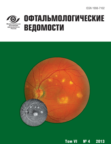
- Выпуск: Том 6, № 4 (2013)
- Страницы: 55-59
- Раздел: Статьи
- Статья получена: 24.06.2015
- Статья опубликована: 15.12.2013
- URL: https://journals.eco-vector.com/ov/article/view/366
- DOI: https://doi.org/10.17816/OV2013455-59
- ID: 366

Цитировать
Полный текст

Аннотация
Врождённая гипертрофия пигментного эпителия - это доброкачественное пигментированное поражение в заднем полюсе, имеющее характерный вид. Оно плоское, округлое, имеет чёткие контуры; обычно чёрного цвета, с внутренними беспигментными лакунами, но может быть совершенно лишенным пигмента. В течение многих лет очаги поражения могут несколько увеличиваться, их пигментация способна меняться. Несмотря на то, что злокачественная трансформация встречается исключительно редко, обследования следует проводить ежегодно.
Ключевые слова
Список литературы
- Reese A. B., Jones I.S. Benign melanomas of the retinal pigment epithelium // Am J. Ophthalmology. - 1956. - Vol. 42. - P. 207.
- Buettner H. Congenital hypertrophy of the retinal pigment epithelium (RPE) A nontumorous lesion // Mod Probl. Ophthalmol. - 1974. - Vol. 12. - P. 528.
- Shields J. A., Augsburger J. J., Brown G. C., Stephens R. F. The differential diagnosis of posterior uveal melanoma // Ophthalmology. - 1980. - Vol. 87. - P. 518-522.
- Coleman P. Barnard N. A. Congenital hypertrophy of the retinal pigment epithelium: prevalence and ocular features in the optometric population // Ophthalmic Physiol Opt. - 2007. - Vol. 27, N 6. - P. 547-555.
- Augsbuger J. J., Henson G. L., Hershbergeer V. S. Trichopoupos N. Topographical distribution of typical unifocal congenital hypertrophy of retinal pigment epithelium 2006 Graefes // Arch. Clin. Exp. Ophthal. - 2006. - Vol. 224. - P. 1412-1414.
- Buettner H. Congenital hypertrophy of the retinal pigment epithelium // Am. J. Ophthal. - 1975. - Vol. 79. - P. 177-189.
- Shields C. L., Mashayekhi A., Ho T. et al. Solitary congenital hypertrophy of the retinal pigment epithelium. Clinical features and frequency of enlargement in 330 patients // Ophthalmology. - 2003. - Vol. 110. - P. 1968-1976.
- Chamot L., Zografos L., Klainguti G. Fundus changes associated with congenital hypertrophy of the retinal pigment epithelium // AM J. Ophthal. - 1993. - Vol. 115. - P. 154-161.
- Shields J. A., Shields C. L., Singh A. D. Acquired tumors arising from congenital hypertrophy of the retinal pigment epithelium // Arch. Ophthalmol. - 2000. - Vol. 118. - P. 637-641.
- Lloyd W. C., Eagle R. C., Shields J. A. et al. Congential Hypertrophy of the Pigment Epithelium Electron Microscopic and morphometric Observations // Ophthalmology. - 1990. - Vol. 97. - P. 1052-1060.
- Cleary P. E., Gregor Z., Bird A. C. Retinal vascular changes in congenital hypertrophy of the retinal pigment epithelium // Brit. J. Ophthal. - 1976. - Vol. 60. - P. 499-503
- Cohen S. Y., Quentel G., Guiberteau B., Coscas G. J. Retinal vascular changes in congenital hypertrophy of the retinal pigment epithelium. Ophthalmology. - 1993. - Vol. 100, N 4. - P. 471-474.
- Shields C. L., Pirondini C., Bianciotto C., Harmon S. A., Shields J. A. Autofluorescence of congenital hypertrophy of the retinal pigment epithelium // Retina. - 2007. - Vol. 27, N 8. - P. 1097-1100.
- Shields C. L., Materin M. A., Walker C., Marr B. P., Shields J. A. Photoreceptor loss overlying congenital hypertrophy of the retinal pigment epithelium by optical coherence tomography // Ophthalmology. - 2006. - Vol. 113, N 4. - P. 661-665.
- Sorsby A. Congenital coloboma of the macula // Br. J Ophthal. - 1935. - Vol. 19. - P. 65-90.
Дополнительные файлы

