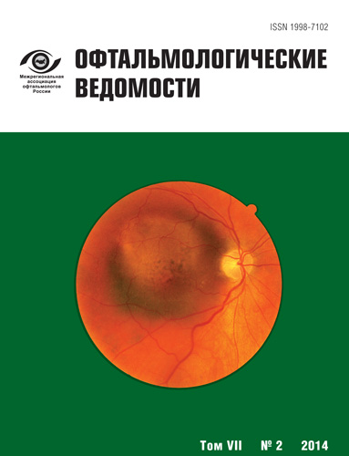
Optical coherence tomography: how it all began, and present-time diagnostic capabilities
- 作者: Astakhov Y.S.1, Belekhova S.G.1
-
隶属关系:
- Pavlov First Saint Petersburg State Medical University
- 期: 卷 7, 编号 2 (2014)
- 页面: 60-68
- 栏目: Articles
- ##submission.dateSubmitted##: 24.06.2015
- ##submission.datePublished##: 15.06.2014
- URL: https://journals.eco-vector.com/ov/article/view/417
- DOI: https://doi.org/10.17816/OV2014260-68
- ID: 417
如何引用文章
全文:
详细
The optical coherence tomography is a modern method to assist the ophthalmologist examine the eye fundus. Tomographs have a very high resolution and give the ophthalmologist a in real-time mode in vivo a detailed examination of retinal, optic nerve and choroidal structures. A continual improvement of this technique offers great opportunities and is not only of scientific but also of practical interest.
作者简介
Yuriy Astakhov
Pavlov First Saint Petersburg State Medical University
Email: astakhov73@mail.ru
MD, doctor of medical science, professor, head of the department of ophthalmology
Svetlana Belekhova
Pavlov First Saint Petersburg State Medical University
Email: beleksv@yandex.ru
Postgraduate student. Department of Ophthalmology
参考
- Васильев В. Н., Гуров И. П. Сравнительный анализ методов оптической когерентной томографии. Известия высших учебных заведений. Приборостроение. 2007; N 7: 30-40.
- Гуров И. П., Козлов С. А. ред. Проблемы когерентной и нелинейной оптики. СПбГУ ИТМО. СПб.; 2004.
- Кальянов А. Л., Лычагов В. В., Лякин Д. В., Перепелицына О. А., Рябухо В. П. Оптическая низкокогерентная интерферометрия и томография. Специальный оптический практикум. 2009; 86.
- Ландсберг Г. С. Оптика. М.: ФИЗМАТЛИТ; 2003.
- Ayton L. N., Guymer R. H., Luu C. D. Choroidal thickness profiles in retinitis pigmentosa. Clin. Experiment Ophthalmol. 2013; 41 (4): 396-403.
- Carl Zeiss Meditec Announces Installation of 6 000th Stratus OCT. Carl Zeiss Meditec, Inc. Dublin, CA. 2006.
- Chung S. E., Kang S. E., Lee J. H., Kim Y. T. Choroidal thickness in polypoidal choroidal vasculopathy and exudative age-related macular degeneration. Ophthalmology. 2011; 118 (5): 840-5.
- Cirrus Hd-Oct Record Number of 10000 Installations and Prestigious Award for Oct Inventors. Carl Zeiss Meditec, Inc., Dublin, CA; 2012.
- Dhoot D. S., Huo S., Yuan A., Xu D., Srivistava S., Ehlers J. P. et al. Evaluation of choroidal thickness in retinitis pigmentosa using enhanced depth imaging optical coherence tomography. Br. J. Ophthalmol. 2012; 97: 66-9.
- Ferguson R. D., Hammer D. X., Paunescu L. A., Beaton S., Schu-man J. S. Tracking optical coherence tomography. Opt. Lett. 2004; 29 (18): 2139-41.
- Forte R., Cennamo G. L., Finelli M. L., Crecchio G. Comparison of Time Domain Stratus OCT and Spectral Domain SLO/OCT for Assessment of Macular Thickness and Volume. Eye. 2009; 23 (11): 2071-8.
- Fujiwara T., Imamura Y., Margolis R., Slakter J. S. et al. Enhanced depth imaging optical coherence tomography of the choroid in highly myopic eyes. Am. J. Ophthalmol. 2009; 148 (3):445-50.
- Garas A., Vargha P., Hollo G. Diagnostic accuracy of nerve fibre layer, macular thickness and optic disc measurements made with the RTVue-100 optical coherencetomograph to detect glaucoma. Eye. 2011; 25: 57-65.
- Geitzenauer W., Hitzenberger C. K., Schmidt-Erfurth U. M. Retinal optical coherence tomography: past, present and future perspectives. Br. J. Ophthalmol. 2011; 95 (2): 171-7.
- Hee M. R., Izatt J. A., Swanson E.A, Huang D., Schuman J. S., Lin C. P., Puliafito C. A., Fujimoto J. G. Optical coherence tomography of the human retina. Arch.Ophthalmol. 1995; 113: 325-32.
- Huang D., Swanson E. A., Lin C. P., Schuman J. S., Stinson W. G., Chang W., Hee M. R., Flotte T., Gregory K., Puliafito C. A., Fujimoto J. G. Optical coherence tomography. Science. 1991; 254: 1178-81.
- Ikuno Y., Kawaguchi K., Yasuno Y., Nouchi T. Choroidal Thickness in Healthy Japanese Subjects. Invest. Ophthalmol. Vis. Sci. 2010; 51 (4): 2173-6.
- Imamura Y., Fujiwara T., Margolis R., Spaide R. F. Enhanced depth imaging optical coherence tomography of the choroid in central serous chorioretinopathy. Retina. 2009; 28: 1469-73.
- Izatt J. A., Hee M. R., Swanson E. A., Lin C. P., Huang D., Schuman J. S., Puliafito C. A., Fujimoto J. G. Micrometer-scale resolution imaging of the anterior eye in vivo with optical coherence tomography. Arch. Ophthalmol. 1994; 11: 1584-9.
- Jonas J. B., Forster T. M., Steinmetz P., Schlichtenbrede F. C., Harder B. C. Choroidal thickness in age-related macular degeneration. Retina. 2014; 34(6): 1149-55.
- Khanifar A. A., Parlitsis G. J., Ehrlich J. R. et al. Retinal nerve fiber layer evaluation in multiple sclerosis with spectral domain optical coherence tomography. Clinical Ophthalmology. 2010; 4: 10071013.
- Kim S. W., Oh J., Kwon S. S., Yoo J., Huh K. Comparison of choroidal thickness among patients with healthy eyes, early age-related maculopathy, neovascular age-related macular degeneration, central serous chorioretinopathy, and polypoidalchoroidal vasculopathy. Retina. 2011; 31: 1904-1911.
- Leung C. K., Cheung C. Y., Weinreb R. N., Lee G. et al. Comparison of Macular Thickness Measurements between Time Domain and Spectral Domain Optical Coherence Tomography Invest. Ophthalmol. Vis. Sci. 2008; 49 (11): 4893-7.
- Manjunath V., Fujimoto J. G., Duker J. S. Cirrus HD-OCT high definition imaging is another tool available for visualization of the choroid and provides agreement with the finding that the choroidal thickness is increased in central serous chorioretinopathy in comparison to normal eyes. Retina. 2010; 30 (8): 1320-1.
- Manjunath V., Goren J., Fujimoto J. G., Duker J. S. Analysis of choroidal thickness in age-related macular degeneration using spectral-domain optical coherence tomography. Am. J. Ophthalmol. 2011; 152: 663-8.
- Manjunath V., Taha M., Fujimoto J. G., Duker J. S. Choroidal thickness in normal eyes measured using Cirrus HD optical coherence tomography. Am. J. Ophthalmol. 2010; 150 (3): 325-31.
- Margolis R., Spaide R. F. A pilot study of enhanced depth imaging optical coherence tomography of the choroid in normal eyes. Am. J. Ophthalmol. 2009; 147 (5): 811-5.
- Martinez-Conde S., Macknik S. L., Hubel D. H. The role of fixational eye movements in visual perception. Nat. Rev. Neurosci. 2004; 5 (3): 229-40.
- Maruko I., Iida T., Sugano Y., Ojima A., Ogasawara M., Spaide R. F. Subfovealchoroidal thickness after treatment of central serous chorioretinopathy. Ophthalmology. 2010; 117 (9): 1792-9.
- McCourt E. A., Cadena B. C., Barnett C. J., Ciardella A. P. et al. Measurement of subfovealchoroidal thickness using spectral domain optical coherence tomography. Ophthalmic Surg. Lasers Imaging. 2010; 41: 28-33.
- Park H. Y., Park C. K. Diagnostic capability of lamina cribrosa thickness by enhanced depth imaging and factors affecting thickness in patients with glaucoma. Ophthalmology. 2013; 120 (4): 745-52.
- Povazay B., Bizheva K., Hermann B. et al. Enhanced visualization of choroidal vessels using ultrahigh resolution ophthalmic OCT at 1050 nm. Opt. Express. 2003; 11 (17): 1980-1986.
- Povazay B., Hermann B., Unterhuber A. et al. Three-dimensional optical coherence tomography at 1050 nm versus 800 nm in retinal pathologies: enhanced performance and choroidal penetration in cataract patients. J. Biomed Opt. 2007; 12 (4).
- Puliafito C. A., Hee M. R., Lin C. P. et al. Imaging of macular diseases with optical coherence tomography. Ophthalmology. 1995; 102: 217-29.
- Schuman J. S., Puliafito C. A., Fujimoto J. G. et al. Optical Coherence Tomography of Ocular Diseases 2nd ed. Thorofare, NJ: SLACK, Inc. 2004.
- Spaide R. F. Enhanced depth imaging optical coherence tomography of retinal pigment epithelial detachment in age-related macular degeneration. Am. J. Ophthalmol. 2009; 147 (4): 644-52.
- Spaide R. F., Koizumi H., Pozzoni M. C. Enhanced depth imaging spectral-domain optical coherence tomography. Am. J. Ophthalmol. 2008; 146 (4): 496-500.
- Swanson E. A., Izatt J. A., Hee M. R., Huang D., Lin C. P., Schu-man J. S., Puliafito C. A., Fujimoto J. G. In vivo retinal imaging by optical coherence tomography. Optics Letters. 1993; 18 (21): 1864-6.
- Tan O., Chopra V., Tzu-Hui A. L. Detection of Macular Ganglion Cell Loss in Glaucoma by Fourier-Domain Optical Coherence Tomography. Ophthalmology. 2009; 116: 2305-14.
- Vienola K. V., Braaf B., Sheehy C. K., Yang Q. et al. Real-time eye motion compensation for OCT imaging with tracking SLO. Biomed Opt Express. 2012; 3 (11): 2950-63.
- Vizzeri G., Weinreb R. N., Gonzalez-Garcia A. O., Bowd C. et al. Agreement between spectral-domain and time-domain OCT for measuring RNFL thickness. Br. J. Ophthalmol. 2009; 93: 775-8.
- Yannuzzi L. A., Freund K. B., Goldbaum M., Scassellati-Sforzolini B., Guyer D. R., Spaide R. F. et al. Polypoidal choroidal vasculopathy masquerading as central serous chorioretinopathy. Ophthalmology. 2000; 107: 767-77.
- You J. Y., Park S. C., Su D., Teng C. C., Liebmann J. M., Ritch R. Focal lamina cribrosa defects associated with glaucomatous rim thinning and acquired pits. JAMA Ophthalmol. 2013; 131 (3): 314-20.
补充文件


