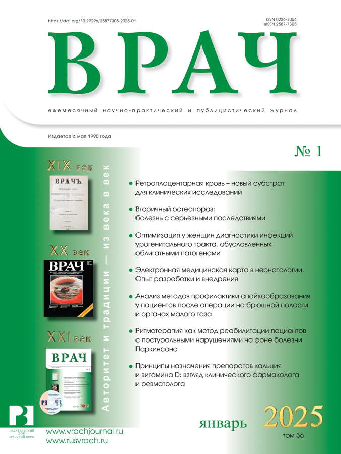
Secondary osteoporosis: a disease with serious consequences
- Authors: Sergeeva-Kondrachenko M.Y.1, Terina N.А.1
-
Affiliations:
- Penza Institute for Postgraduate Medical Education
- Issue: Vol 36, No 1 (2025)
- Pages: 9-16
- Section: Lecture
- URL: https://journals.eco-vector.com/0236-3054/article/view/676815
- DOI: https://doi.org/10.29296/25877305-2025-01-02
- ID: 676815
Cite item
Abstract
Secondary osteoporosis (OP) develops as a result of somatic pathologies (endocrine, genetic diseases, kidney damage, gastrointestinal tract, etc.), lifestyle habits or medications. Any patient with suspected secondary OP must undergo a series of laboratory tests (full clinical blood test, biochemical blood test, blood test for 25-hydroxyvitamin D, parathyroid hormone, etc.). The “gold standard” for instrumental diagnosis of OP is dual-energy X-ray densitometry of the lumbar spine and proximal femur to assess bone mineral density.
Treatment of secondary OP is etiological, aimed at identifying and eliminating the underlying cause of the disease, which usually involves discontinuation of medications (if possible) and correction of modifiable risk factors.
If it is impossible to influence the cause of secondary OP, pathogenetic therapy is used, including antiresorptive drugs, agents that enhance bone formation, and monoclonal antibodies. It is important to remember that the effectiveness of treatment of OP is influenced by indicators of phosphorus-calcium metabolism, therefore, before starting pathogenetic therapy, it is necessary to determine the levels of calcium, phosphorus and 25-hydroxyvitamin D in the blood, bring these indicators to normal values, and then continue taking these drugs along with pathogenetic therapy. To do this, they recommend a diet balanced in calcium, phosphorus and proteins, and the prescription of vitamin D supplements and calcium salts. One of the effective means for normalizing phosphorus-calcium metabolism is the drug Osteomed Forte.
Full Text
About the authors
M. Yu. Sergeeva-Kondrachenko
Penza Institute for Postgraduate Medical Education
Email: marserkon@mail.ru
MD
Russian Federation, PenzaN. А. Terina
Penza Institute for Postgraduate Medical Education
Author for correspondence.
Email: marserkon@mail.ru
Russian Federation, Penza
References
- Aspray T.J., Hill T.R. Osteoporosis and the Ageing Skeleton. Subcell Biochem. 2019; 91: 453–76. doi: 10.1007/978-981-13-3681-2_16
- Foessl I., Dimai H.P., Obermayer-Pietsch B. Long-term and sequential treatment for osteoporosis. Nat Rev Endocrinol. 2023; 19 (9): 520–33. doi: 10.1038/s41574-023-00866-9
- LeBoff M.S., Greenspan S.L., Insogna K.L. et al. The clinician's guide to prevention and treatment of osteoporosis. Osteoporos Int. 2022; 33 (10): 2049–102. doi: 10.1007/s00198-021-05900-y
- Petrie J.R., Marso S.P., Bain S.C. et al. LEADER-4: blood pressure control in patients with type 2 diabetes and high cardiovascular risk: baseline data from the leader randomized trial. J Hypertension. 2016; 34 (6): 1140–50. doi: 10.1097/HJH.0000000000000890
- Sheu A., Diamond T. Secondary osteoporosis. Aust Prescr. 2016; 39 (3): 85–7. doi: 10.18773/austprescr.2016.038
- Sobh M.M., Abdalbary M., Elnagar S. et al. Secondary Osteoporosis and Metabolic Bone Diseases. J Clin Med. 2022; 11: 2382. doi: 10.3390/jcm11092382
- Strukov V.I., Kislov A.I., Eremina N.V. et al. The use of bone tissue non-steroid anabolizators in treatment of osteoporosis. Research Journal of Pharmacy and Technology. 2019; 12 (5): 2195–9. doi: 10.5958/0974-360X.2019.00366.4
- Xiao P.L., Cui A.Y., Hsu C.J. et al. Global, regional prevalence, and risk factors of osteoporosis according to the World Health Organization diagnostic criteria: a systematic review and meta-analysis. Osteoporos Int. 2022; 33 (10): 2137–53. doi: 10.1007/s00198-022-06454-3
- Белая Ж.Е., Белова К.Ю., Бирюкова Е.В. и др. Федеральные клинические рекомендации по диагностике, лечению и профилактике остеопороза. Остеопороз и остеопатии. 2021; 2: 4–47 [Belaya Zh.E., Belova K.Yu., Biryukova E.V. et al. Federal clinical guidelines for the diagnosis, treatment and prevention of osteoporosis. Osteoporoz i osteopatii. 2021; 2: 4–47 (in Russ.)]. doi: 10.14341/osteo12930
- Кондраченко М.Ю. Влияние Кальция Д3-Никомед на минеральную плотность костной ткани у больных сахарным диабетом I типа. Пермский медицинский журнал. 2006; 23 (1): 40–8 [Kondrachenko M.Yu. The effect of Calcium D3-Nicomed on bone mineral density in patients with type I diabetes mellitus. Permskij medicinskij zhurnal. 2006; 23 (1): 40–8 (in Russ.)].
- Кондраченко М.Ю. Минеральная плотность костной ткани при сахарном диабете I типа разной продолжительности заболевания и степени компенсации углеводного обмена. Вестник Волгоградского государственного медицинского университета. 2006; 2 (18): 51–5 [Kondratenko M.Y. Bone mineral density in type I diabetes mellitus of different disease duration and degree of compensation of carbohydrate metabolism. Vestnik Volgogradskogo gosudarstvennogo medicinskogo universiteta. 2006; 2 (18): 51–5 (in Russ.)].
- Сергеева-Кондраченко М.Ю. Постменопаузальный остеопороз и сахарный диабет 2 типа: отношения с угрозой для жизни. В сб.: Актуальные вопросы диагностики, лечения и реабилитации больных. Мат-лы XX Межрег. научно-практ. конф. Пенза, 2020; с. 182–4 [Sergeeva-Kondrachenko M.Y. Postmenopausal osteoporosis and type 2 diabetes mellitus: life-threatening relationships. In the collection: Topical issues of diagnosis, treatment and rehabilitation of patients. Materials of the XX Interregional scientific and practical conference. Penza, 2020; pp. 182–4 (in Russ.)].
- Струков В., Елистратов Д., Балыкова Л. и др. Влияние Остеомеда Форте на гормональный статус и течение остеопороза у женщин с дефицитом андрогенов в постменопаузе. Врач. 2015; 3: 28–32 [Strukov V., Elistratov D., Balykova L. et al. The effect of Osteomed Forte on hormonal status and the course of osteoporosis in postmenopausal women with androgen deficiency. Vrach. 2015; 3: 28–32 (in Russ.)].
- Струков В.И., Сергеева-Кондраченко М.Ю., Виноградова О.П. и др. Коморбидный пациент с остеопорозом на приеме у врача. Какие факторы необходимо учитывать в подборе терапии. Медицинский алфавит. 2023; 18: 34–8 [Strukov V.I., Sergeeva-Kondrachenko M.Yu., Vinogradova O.P. et al. Comorbid patient with osteoporosis at doctor's appointment. What factors should be considered in selection of therapy. Medical alphabet. 2023; 18: 34–8 (in Russ.)]. doi: 10.33667/2078-5631-2023-18-34-38
- Струков В.И., Сергеева-Кондраченко М. Ю., Струкова-Джоунс О.В. и др. Актуальные проблемы остеопороза: монография. Ростра, 2009; 342 с. [Strukov V.I., Sergeeva-Kondrachenko M. Yu., Strukova-Jones O.V. et al. Current problems of osteoporosis: a monograph. Rostra, 2009; 342 p. (in Russ.)].
- Джоунс О., Струков В., Кислов А. и др. Коморбидный остеопороз: проблемы и новые возможности терапии (ч. 1). Врач. 2017; 10: 23–6 [Jones O., Strukov V., Kislov A. et al. Comorbid osteoporosis: problems and new possibilities of therapy (Part 1). Vrach. 2017; 10: 23–6 (in Russ.)].
- Джоунс О., Струков В., Кислов А. и др. Коморбидный остеопороз: проблемы и новые возможности терапии (ч. 2). Врач. 2017; 11: 25–8 [Jones O., Strukov V., Kislov A. et al. Comorbid osteoporosis: problems and new possibilities of therapy (Part 2). Vrach. 2017; 11: 25–8 (in Russ.)].
- Хабибулина М.М., Шамилов М.Д., Елистратов Д.Г. Эффективная фармакотерапия остеопороза в перименопаузе. Врач. 2024; 35 (11): 38–40 [Khabibulina M., Shamilov M., Elistratov D. Effective pharmacotherapy of osteoporosis in perimenopause. Vrach. 2024; 35 (11): 38–40 (in Russ.)]. doi: 10.29296/25877305-2024-11-07
- Шамилов М.Д., Хабибулина М.М. Остеопороз: диагностика и лечение коморбидных пациентов, персонифицированный подход к терапии. Врач. 2024; 35 (7): 24–9 [Shamilov M., Khabibulina M. Osteoporosis: diagnosis and treatment of comorbid patients, a personalized approach to therapy. Vrach. 2024; 35 (7): 24–9 (in Russ.)]. doi: 10.29296/25877305-2024-07-04
Supplementary files


