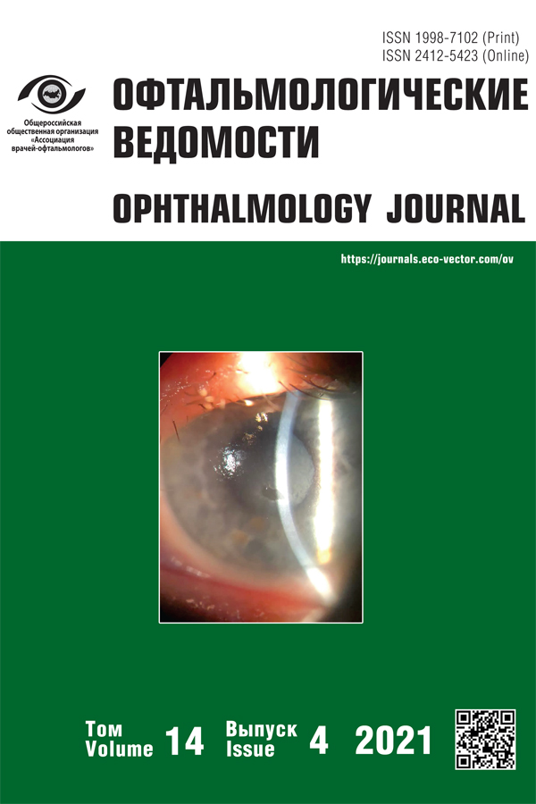
Pathomimia in a practice of an ophthalmologist
- Authors: Potemkin V.V.1,2, Marchenko O.A.2, Goltsman E.V.2, Anikina L.K.1, Gladysheva E.K.1
-
Affiliations:
- Pavlov First St. Petersburg State Medical University
- City Multidiscipline Hospital No. 2
- Issue: Vol 14, No 4 (2021)
- Pages: 73-78
- Section: Case reports
- Submitted: 18.02.2022
- Accepted: 18.02.2022
- Published: 15.12.2021
- URL: https://journals.eco-vector.com/ov/article/view/101088
- DOI: https://doi.org/10.17816/OV101088
- ID: 101088
Cite item
Abstract
Non-suicidal self-inflicted injuries are encountered in the practice of doctors of all specialties. Within the framework of this article, a case of surgical treatment of a female patient with pathomimia and lagophthalmos will be presented.
Full Text
BACKGROUND
Non-suicidal self-inflicted injuries are a variant of auto-aggression without the intention to commit suicide [1, 2]. One of the challenging aspects is establishing a diagnosis because patients often visit not psychiatrists but doctors of other specialties for the treatment of self-inflicted injuries. Doctors of all specialties encounter these conditions, and ophthalmologists are not exempted.
Self-injury to the eye can take many forms, from eye enucleation to superficial damage to the anterior part of the eye. Thus, enucleation [3–6] and damage to the orbit [7–11], ocular surface, and anterior [12–15] and posterior segments of the eyeball [16–20] have been described. One of the most common forms of non-suicidal self-inflicted injuries is skin injury, i.e., pathomimia [21].
This article presents the clinical case of a patient with non-suicidal self-inflicted injuries of the skin of the eyelids and surrounding tissues.
CLINICAL CASE
A 49-year-old female patient was hospitalized on an emergency basis at the Department of Ophthalmology No. 5 of the St. Petersburg City Multidisciplinary Hospital No. 2 because of cicatricial lagophthalmos. Upon admission, she complained of incomplete closure of the right palpebral fissure, a long-term non-healing wound of the soft tissues of the face, accompanied by pain, and a constant foreign body sensation. The patient revealed «silicone residues in the edges of the wound from the cosmetic fillers inserted.» She treated the wound independently, denied having had any injury, and described in detail about how she «removes particles of perceptible silicone by pressing on the wound edges, which induces its release from the wound as a transparent liquid.»
Regarding medical history, the patient repeatedly received various cosmetic injections in the face since the 1990s. The first episode of pain syndrome with tissue necrosis and a long-term non-healing wound was noted in 2004 after rhinoplasty. Medical history was analyzed based on the medical documentation provided by the patient. In 2017, she had surgical treatment of a long-term non-healing wound in the upper third of the left nasolabial fold. In 2019, she underwent surgical treatment of a long-term non-healing wound in the forehead, and in 2019, she refused surgical treatment of a long-term penetrating soft tissue defect of the nasal dorsum and septum on the left.
One of the most challenging aspects of this clinical case was the patient’s mental status. According to the patient, she consulted with psychiatrists many times, and no psychiatric pathology was detected. However, in 2019, during one of the hospitalizations, the patient was consulted by a psychiatrist and was diagnosed with developed nosomania.
During a conversation with the patient, the patient constantly touched the wounds, compressing their edges and squeezing out the sanious discharge, and lifted the skin flap without experiencing pain. She considered the sanious discharge and the granular surface of the wound as foreign bodies, the remains of fillers. The patient’s complaints were systematized in accordance with her concept of the disease; she had no criticism of her condition.
An objective examination of the facial skin surface in the region of the eyelid, eyebrow, and forehead on the right revealed a penetrating wedge-shaped skin defect, 6.0 × 4.0 cm in size, a chronic fibrinous-granulating wound with signs of epithelialization, and a 3-mm lagophthalmos (Fig. 1). The wound had smooth and tucked edges with small hemorrhages (fibrin). There were pockets on the sides of the wound. At the wound bottom, there were pale granulation tissues, with mild contact bleeding, without discharge, including pus. There were multiple transplanted skin flaps of irregular shape on the face and facial asymmetry due to the cicatricial deformity of the soft tissues in the left nasolabial fold, nasal dorsum, and glabella (Fig. 2).
Fig. 1. Appearance of the wound before surgery, granulation tissue in the wound bed, profound pockets at the edges, incomplete eyelid closure (3 mm) / Рис. 1. Внешний вид раны до хирургического лечения, дно раны представлено грануляционной тканью, по краям имеются глубокие карманы, несмыкание глазной щели составляет 3 мм
Fig. 2. Multiple transplanted skin flaps, cicatricial deformity of the bridge of the nose / Рис. 2. Множественные пересаженные кожные лоскуты, рубцовая деформация спинки носа
Despite clear signs of non-suicidal self-inflicted injuries in the patient, surgical treatment was required for cicatricial lagophthalmos. Surgical treatment was preceded by spiral computed tomography of the orbits to assess the state of tissues in the proposed surgical site and exclude the presence of silicone. Data on the presence of silicone in both the orbit and surrounding tissues have not been received. In the upper eyelid, an air bubble was detected in the skin pocket at the edge of the wound (Fig. 3, a, b). Compared with air, silicone has a much higher, close to bone, density.
Fig. 3. Spiral computed tomogram of orbits. a, b – air bubble at the right of the upper eyelid area / Рис. 3. Спиральная компьютерная томограмма орбит: a, b — пузырёк воздуха в области верхнего века справа
A free skin graft was transplanted from the postauricular area. At the first stage, the skin of the wound edges was excised (Fig. 4, a), and the granulation tissue was removed from the bottom and edges (Fig. 4, b, c). The material was sent for histological examination.
Fig. 4. Stages of surgery: renewal of the wound edges (a) and removal of granulation tissue from the bottom and from the edges (b, c) / Рис. 4. Этапы операции: обновление краёв раны (a), удаление грануляционной ткани со дна и краёв (b, c)
A full-thickness skin graft of approximately 6.0 × 4.0 cm in size was isolated with a scalpel in the postauricular area and excised (Fig. 5, a), and hemostasis was performed (Fig. 5, b). The wound edges were mobilized and sutured with interrupted sutures (Fig. 5, c).
Fig. 5. Stages of surgery. Behind-the-ear area: skin graft is removed (a), hemostasis (b), wound sutured with interrupted sutures (c) / Рис. 5. Этапы операции. Заушная область: кожный трансплантат изъят (a), гемостаз (b), рана ушита узловыми швами (с)
A full-thickness free skin graft was placed according to the defect size and fixed with guiding interrupted sutures. Then, the wound edges were adapted along the entire perimeter with a continuous suture (Fig. 6, a, b).
Fig. 6. Defect closure with a free skin flap (a), fixation with interrupted and running sutures (b) / Рис. 6. Закрытие дефекта свободным кожным лоскутом (a), фиксация узловыми и непрерывным швами (b)
Postoperatively, a pressure bandage was applied. The patient was followed up for a week. At discharge, the engraftment of the flap was complete, the edges were fully adapted, and no diastasis or necrosis was noted. The wound in the postauricular area was also consistent, without signs of inflammation.
At the control examination on day 14, the wound edges were adapted, and the flap engraftment was complete, with no signs of rejection. Postoperative sutures were removed.
One month after the surgery, the patient revisited the department with complaints of a penetrating skin wound and presence of foci of necrosis along the edges (Fig. 7). According to the patient, in the area of the postoperative wound, she again found silicone remains, and she tried to remove them. The patient again underwent surgical treatment similar to the previous one, taking into account the presence of lagophthalmos (Figs. 7 and 8).
Fig. 7. Appearance of the wound. One month after surgery / Рис. 7. Внешний вид раны через месяц после операции
Fig. 8. Repeated surgical treatment: a – removal of granulation tissue and renewal of the wound edges; b – taking a skin flap from the behind-the-ear area; c – fixation of the skin flap /
Рис. 8. Повторное хирургическое лечение: a — удаление грануляционной ткани и обновление краёв раны; b — взятие кожного лоскута из заушной области; c — фиксация кожного лоскута
Unfortunately, the outcome of the repeated intervention was similar to the previous one. For the third time, the patient was denied surgical treatment, since the non-closure of the palpebral fissure was less than 1 mm and there were no signs of corneal xerosis.
The case presents that surgical treatment of patients with non-suicidal self-inflicted injuries does not seem to be adequately effective without prior psychiatric correction with medications and/or therapy. The patient was repeatedly referred for a consultation to a neuropsychiatric dispensary to correct her condition.
CONCLUSION
Non-suicidal self-inflicted injuries cause clinically significant suffering and problems in all spheres of life; thus, timely provision of specialized psychiatric care is an integral part of achieving success in the treatment of such patients.
ADDITIONAL INFORMATION
Author contributions. All authors confirm that their authorship complies with the ICMJE criteria. All authors have made a significant contribution to the development of the concept, research, and preparation of the article, read, and approved the final version before its publication.
Funding. The study had no external funding.
Conflict of interest. The authors declare no conflict of interest.
Informed consent to publication. The authors obtained the written consent of the patient’s legal representatives for the publication of medical data and photographs.
About the authors
Vitalii V. Potemkin
Pavlov First St. Petersburg State Medical University; City Multidiscipline Hospital No. 2
Email: potem@inbox.ru
ORCID iD: 0000-0001-7807-9036
SPIN-code: 3132-9163
Cand. Sci. (Med.), Head of the Microsurgical Department (eyes) No. 5
Russian Federation, 6-8, L’va Tolstogo st., Saint Petersburg, 197022; Saint PetersburgOlga A. Marchenko
City Multidiscipline Hospital No. 2
Email: oamarchenko@yandex.ru
Ophthalmologist
Russian Federation, Saint PetersburgElena V. Goltsman
City Multidiscipline Hospital No. 2
Email: ageeva_elena@inbox.ru
ORCID iD: 0000-0002-2568-9305
Ophthalmologist
Russian Federation, Saint PetersburgLiliya K. Anikina
Pavlov First St. Petersburg State Medical University
Email: lily-sai@yandex.ru
ORCID iD: 0000-0001-8794-0457
Postgradeate student
Russian Federation, 6-8, L’va Tolstogo st., Saint Petersburg, 197022Ekaterina K. Gladysheva
Pavlov First St. Petersburg State Medical University
Author for correspondence.
Email: gladysheva.e.k@ya.ru
ORCID iD: 0000-0001-9186-0994
Resident of the Department of Ophthalmology
Russian Federation, 6-8, L’va Tolstogo st., Saint Petersburg, 197022References
- Sergeev II, Levina SD. Nesuitsidal’nye samopovrezhdeniya pri rasstroistvakh shizofrenicheskogo spektra. Moscow: Tsifrovichok, 2009. 171 p. (In Russ.)
- Meszaros G, Horvath LO, Balazs J. Selfinjury and externalizing pathology: a systematic literature review. BMC Psychiatry. 2017;17(1):160. doi: 10.1186/s12888-017-1326-y
- Khan JA, Buescher L, Ide CH, Pettigrove B. Medical management of self-enucleation. Arch Ophthalmol. 1985;103(3):386–389. doi: 10.1001/archopht.1985.01050030082027
- Field HL, Waldfogel S. Severe ocular self-injury. Gen Hosp Psychiatry. 1995;17(3):224–227. doi: 10.1016/0163-8343(95)00031-l
- Jones NP Self-enucleation and psychosis. Br J Ophthalmol. 1990;74(9):571–573. doi: 10.1136/bjo.74.9.571
- Stannard K, Leonard T, Holder G, Shilling J. Oedipism reviewed: a case of bilateral ocular self-mutilation. Br J Ophthalmol. 1984;68(4):276–280. doi: 10.1136/bjo.68.4.276
- Tapper CM, Bland RC, Danyluk L. Self-inflicted eye injuries and self-inflicted blindness. J Nerv Ment Dis. 1979;167(5):311–314. doi: 10.1097/00005053-197905000-00008
- Lasky JB, Epley KD, Karesh JW. Household objects as a cause of self-inflicted orbital apex syndrome. J Trauma. 1997;42(3):555–558. doi: 10.1097/00005373-199703000-00030
- Bowen DI. Self-inflicted orbitocranial injury with a plastic ballpoint pen. Br J Ophthalmol. 1971;55(6);427–430. doi: 10.1136/bjo.55.6.427
- Albert DM, Burns WP, Scheie HG. Severe orbitocranial foreign-body injury. Am J Ophthalmol. 1965;60(6):1109–1111. doi: 10.1016/0002-9394(65)92825-4
- Shuttleworth GN, Galloway PH. Ocular air-gun injury: 19 cases. J Roy Soc Med. 2001;94(8):396–399. doi: 10.1177/014107680109400806
- Yang HK, Brown GC, Magargal LE. Self-inflicted ocular mutilation. Am J Ophthalmol. 1981;91(5):658–663. doi: 10.1016/0002-9394(81)90070-2
- Palmowski A, Heinz G, Ruprecht KW. Self-inflicted injuries of the eye: differential diagnosis of self-inflicted lacerating corneal injury. Klin Monatsbl Augenheilkd. 1994;204(1):30–32. doi: 10.1055/s-2008-1035498
- Kennedy BL, Feldmann TB. Self-inflicted eye injuries: case presentations and a literature review. Hosp Community Psychiatry. 1994;45(5):470–474. doi: 10.1176/ps.45.5.470
- Chern KC, Meisler DM, Wilhelmus KR, et al. Corneal anesthetic abuse and Candida keratitis. Ophthalmology. 1996;103(1):37–40. doi: 10.1016/s0161-6420(96)30735-5
- Brown R, al-Bachari MA, Kambhampati KK. Self-inflicted eye injuries. Br J Ophthalmol. 1991;75(8):496–498. doi: 10.1136/bjo.75.8.496
- Ashkenazi I, Shahar E, Brand N, et al. Self-inflicted ocular mutilation in the pediatric age group. Acta Paediatr. 1992;81(8):649–651. doi: 10.1111/j.1651-2227.1992.tb12323.x
- Noel LP, Clarke WN. Self-inflicted ocular injuries in children. Am J Ophthalmol. 1982;94(5):630–633. doi: 10.1016/0002-9394(82)90008-3
- Stinnett JL, Hollender MH. Compulsive self-mutilation. J Nerv Ment Dis. 1970;150(5):371–375. doi: 10.1097/00005053-197005000-00005
- Blackmon DM, Calvert HM, Henry PM, Westfall CT. Bacillus cereus endophthalmitis secondary to self-inflicted periocular injection. Arch Ophthalmol. 2000;118(11):1585–1586. doi: 10.1001/archopht.118.11.1585
- Grebenyuk VN. Patomimii (obzor literatury). Vestnik dermatologii. 1977;(9):28–32. (In Russ.)
Supplementary files


