


Cases of intraocular lens opacification in pseudophakic eyes: analysis of the results of microstructural studies
- Authors: Riks I.A.1, Astakhov S.Y.1, Ivankova E.М.2, Kuzmina I.E.3, Papanyan S.S.1, Boutaba R.1, Ezugbaya M.B.3, Akopov E.L.3
-
Affiliations:
- Academician I.P. Pavlov First St. Petersburg State Medical University of the Ministry of Healthcare of the Russia
- Institute of Macromolecular Compounds Russian Academy of Sciences
- Academican I.P. Pavlov First St. Petersburg State Medical University of the Ministry of Healthcare of the Russian Federation
- Issue: Vol 13, No 3 (2020)
- Pages: 21-28
- Section: Original study articles
- Submitted: 08.08.2020
- Accepted: 13.10.2020
- Published: 08.01.2021
- URL: https://journals.eco-vector.com/ov/article/view/41836
- DOI: https://doi.org/10.17816/OV41836
- ID: 41836

Cite item

Abstract
Relevance. Currently, all over the world, during cataract surgeries, a huge number of intraocular lenses (IOLs) made of different materials are implanted. Alongside with the development of modern IOL materials and designs, publications about their opacities appear. The nature and the localization of IOL opacities mainly depend on the properties of the material out of which the lens is made. Polymethyl methacrylate (PMMA) currently rarely used to manufacture IOLs, tends to cloud in the optical center due to structural breakdown, forming “snowflake”-like cracks. Opacities of acrylic IOLs depend on the degree of hydrophilic properties of the material. The deposition of crystalline deposits in the optical zone of hydrophilic acrylic lenses leads to a significant decrease in visual acuity and requires IOL explantation. There is a definite dependence of the occurrence of opacities in hydrophilic acryl on the patient’s concomitant diseases. In hydrophobic acrylic IOLs, vacuoles form, and glistenings occurs. Herewith, visual functions, as a rule, do not suffer.
Purpose: to find out what structural changes in the IOL led to the need to remove them from pseudophakic eyes due to a decrease in visual acuity.
Materials and methods. Four clouded IOLs made from different materials were examined. The lenses were studied using a SUPRA 55VP scanning electron microscope (Carl Zeiss, Germany) using a secondary electron detector. Element distribution maps on the surface and inside the lenses were collected using an X-max 80 mm2 energy dispersive X-ray analysis detector (Oxford Instruments, UK).
Results. A hydrophilic lens with hydrophobic coating became cloudy 5 years after implantation. Hydroxyapatite crystals were found on all parts of the IOL along its surface. In a hydrophobic acrylic IOL, microvacuoles and cavities in the optical center were found using scanning electron microscopy. Two PMMA IOLs underwent self-destruction within 8 years after implantation. Chemical analysis of PMMA lenses did not reveal any inorganic compounds.
Conclusion. One of the complications of IOL implantation is an impairment of their transparency. Factors associated with IOL material and manufacturing, as well as the patient’s comorbidities, can lead to lens opacification at various terms after surgery.
Full Text
INTRODUCTION
Currently, all over the world, during cataract surgeries, millions of intraocular lenses (IOLs) made of different materials are implanted. The intraocular lens (IOL) presence inside the eye could lead to several complications. Special attention should be paid to cases when IOL is to be removed from the eye, and a new one is to be implanted. Alongside with the development of modern IOL materials and designs, publications about their opacities appear. In 2008, a large study of explantation causes of 146 IOLs was performed [1]. On the first place, there were IOL calcification and opacities (65%), on the second one – dislocations (up to 23%), aberrations with hydrophobic acrylic lenses had the same prevalence; a somewhat smaller amount of IOL explantations (21%) were due to incorrect optical power calculation. There are evidences that concomitant diseases (local and systemic ones) may influence the pattern of IOL opacities.
Regardless of the fact that materials with best technical characteristics are used for IOL production, many reports are published on opacities, color changes, and destruction of implanted IOLs [1–4]. Most often, IOLs are manufactured out of polymethyl methacrilate (PMMA), silicon, hydrogel, acryl. All mentioned chemical substances may change more or less depending on the environmental conditions (temperature, medications) or on the fact of their persistence inside the eyeball. What is more, such changes as opacities and color changes may occur before long or several years after the IOL implantation, on average, in 3 years after surgery [5, 6].
In the literature, there are descriptions of “cold opalescence”, characteristic for hydrophobic acrylic IOLs. In a steep increase of the temperature, changes in lens material occur, so called phase splitting, which causes a sudden white hint at implantation into the eye [6]. This effect is reversible – at equalization of the lens temperature with that of the intraocular fluid, acrylic IOL becomes transparent. Lenses most frequently used for implantation are manufactured from acryl with different degree of original substance hydration: modern hydrophilic acryl contains 18–28% of water, hydrophobic one – up to 1%.
Some years ago, hydrophilic IOLs were widely used. This was due, in the first instance, to well-proven production technologies; in the second instance, hydrophilic IOLs cause less patients’ complaints on dysmorphopsia appearance, rather than hydrophobic ones [7]. On the other hand, it became obvious that namely hydrophilic material is most often subject to changes, which are related both to lens material and to concomitant diseases of the patient (glaucoma, diabetes mellitus) that cause the appearance in the anterior chamber fluid of substances not characteristic for it [1–4]. In such patients, it is recommended to implant hydrophobic IOLs, as opacities happen significantly more rarely in them.
Cases of hydrophilic acryl’s blue staining due to material absorption of different dyes (fluorescein, indocyanine green, trypan blue) are well known [6].
In 2015, A. Gamidov et al. [7] proposed a classification of IOL opacities: progressing IOL degradation (destruction); IOL opacity, or its color change; presence of hollow microinclusions buried in the lens; crystalline deposits on the IOL surface. The pattern of such changes depends on the IOL material. Opacities have different distribution area: only on the anterior or posterior surface, or on the whole surface and haptics of the IOL. Sometimes, opacities buried in the IOL optic are found [6].
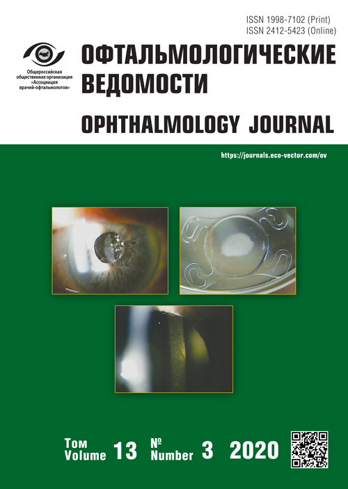
A “glistening” phenomenon is a term, which is common to encounter recently in scientific literature when IOL opacities are described. Sometimes, in Russian articles it is used without translation [7]. This phenomenon occurs in hydrophobic acrylic IOLs due to formation of microvacuoles in the optic part of the lens (Fig. 1). The number of vacuoles may vary, but as a rule, they occupy all the optic part of the lens. The dimension of microvacuoles is 1–20 microns. The problem arises because in IOL production, polymers are used, which after implantation absorb the fluid into their “architectural” structure. Polymers usually take up water at their immersion for a long time in a water medium, and the speed of this process increases with increasing temperatures. For example, if the lens is soaked in warm water, and then the temperature is decreased, water molecules gather in polymer’s cavities. Irregular refraction occurs due to the difference in refraction indices of water and IOL material. In the literature, there are indications on the absence of direct correlation between the number of lens vacuoles and the rationale of given IOL exchange. It is necessary to take into account patients’ complaints of visual acuity decrease and of glistening phenomenon. Only in such case, a question of lens explantation may be raised [8]. It is known that microcavities in hydrophobic acrylic IOLs definitely shape during 3 years after implantation, and the effect of glistening becomes stable [6, 8].

Fig. 1. Biomicroscopy: the phenomenon of “glistenings” in hydrophobic intraocular lens
Рис. 1. Биомикроскопия: феномен «блёсток» в гидрофобной интраокулярной линзе
The opacification of hydrophilic acrylic IOLs is considered to be their major drawback. The data of histochemical, histopathological studies as well as of light electron microscopy proved that opacities are related to precipitation of calcium and calcium phosphate on the surface and/or inside the IOL (Fig. 2).

Fig. 2. Photo of an explanted intraocular lens with calcifications in the optical part
Рис. 2. Фотография эксплантированной интраокулярной линзы с кальцификатами в оптической части
The pathologic calcification of hydrophilic acryl is a multifactorial problem, which includes stages of IOL production and packing, as well as the chemical composition of the patient’s anterior chamber fluid. In hydrophilic acrylic IOLs, opacities form on the surface as crystalline deposits (granules), which may lie as separate groups or coalesce, forming roughnesses. In the literature, such lesions are defined as “dystrophic” calcification. As a rule, these changes occur in patients with concomitant diseases: systemic (diabetes mellitus, arthritis, etc.) or ocular (operated glaucoma, keratoplasty, vitrectomy). In some studies, there are indications that in patients with above-mentioned conditions in the anterior chamber fluid and in the blood increased levels of calcium phosphate are found [3, 4, 7]. Calcium deposits on IOLs are described in cases of postoperative intraocular inflammation, in single-staged combined surgery, and in use of substances introduced into the eye – tissue plasminogen activator, silicone oil, air, or gas [6]. In such cases, opacifications of hydrophilic IOLs occur much earlier, than in “dystrophic” calcification, whereby calcium deposition and its amount may significantly vary. It is known that after “triple” single-staged surgery – phacoemulsification, hydrophilic IOL implantation, posterior lamellar keratoplasty that includes air and/or gas use, – calcification on the IOL’s surface develops, which is always limited by pupil area [9, 10]. To explain precisely these local precipitations of non-organic crystals, further investigations are necessary.
Slowly progressing opacities of “snowflake” type are well known, with multiple cracks in a PMMA IOL looking like a snowflake pattern (Fig. 3). PMMA IOLs were molded using “injection forming”. Such lenses were actively implanted in the Russian Federation until the end of nineties. As a rule, such changes begin after 10 and more years after implantation [11]. As inhomogeneous snowflake-like opacities form always only in the optic part of the IOL, there is a suggestion that ultraviolet irradiation contacting with PMMA steadily destroys this material, forming microdamages with varying directions of cracks.

Fig. 3. Biomicroscopy: polymethyl methacrylate intraocular lens – “snowflake”-like opacities
Рис. 3. Биомикроскопия: интраокулярная линза из полиметилметакрилата — помутнения по типу «снежинка»
The aim of the study – to investigate, which structural changes of IOLs led to the necessity of their removal from pseudophakic eyes associated with visual acuity decrease.
MATERIALS AND METHODS
4 IOLs from 4 patients aged 61–77 years were included in the study. Lenses were explanted because of significant transparency loss and visual acuity decrease: IOL made of hydrophilic acryl with hydrophobic coating, IOL made of hydrophobic acryl, two samples of PMMA IOLs. Surgical lens replacement was performed in the Ophthalmology department of the Academician I.P. Pavlov First St. Petersburg State Medical University. IOLs were examined at the Institute of Macromolecular Compounds, Russian Academy of Sciences. Light electron microscopy examination was carried out with scanning electron microscope Supra 55VP (Carl Zeiss, Germany) using a secondary electron detector. Samples were fixed with special glue on microscope holders and spattered with a thin platinum layer. Maps of element distribution were composed using a detector of energy dispersive X-ray analysis X-max 80 mm2 (Oxford Instruments, Great Britain).
RESULTS
In 2020, first patient included in the present study, applied to the Ophthalmology department of the Academician I.P. Pavlov First St. Petersburg State Medical University complaining of visual acuity decrease on his right eye. Visual acuity was 0.5 (no correction possible); left eye – 1.0. On examination: cornea of the right eye is transparent; average anterior chamber depth, its fluid is transparent; no pathological iris changes; IOL correctly centered, there is a white total opacity in the optic part (Fig. 1), by biomicroscopy, on the IOL surface, there were areas of small coalescing fissures. Left eye – transparent cornea, average anterior chamber depth, its fluid is transparent; no pathological iris changes; IOL transparent and correctly centered. The patient did not have any concomitant systemic disease. In 2014, in this patient, with a several months interval, phacoemulsification surgery with simultaneous implantation of an IOL Oculentis (German production), hydrophilic acrylic IOL with hydrophobic coating, was performed. Surgical procedures were uneventful. In 1 year after surgery, visual acuity of both eyes was 1.0. In 2020, on the right eye, IOL replacement surgery was performed. After explantation, light optic microscopy of the opacified IOL was done. A diffuse opacification of the whole IOL surface was established, including optic part and haptics. White deposits consisted from granules, the coalescence of which caused rough, uneven surface. On microphotographs obtained using electron microscope, one can see round protruding structures up to 5 microns, which coalesce into large “fields”.
Using the detector of energy dispersive X-ray analysis, the chemical composition of white crystalline granules on the IOL was established: calcium hydroxyapatite (Fig. 4). High density of crystals was concentrated in the optic part of the IOL. Small cracks and fissures of the lens could be evaluated in more detail on microphotographs by light microscopy (Fig. 5).

Fig. 4. Microphotography of the explanted intraocular lens Oculentis using a Supra 55VP scanning electron microscope (Carl Zeiss, Germany). Crystals of calcium hydroxyapatite: a – scale 10 μm; b – scale 2 μm
Рис. 4. Микрофотографии эксплантированной интраокулярной линзы Oculentis, полученные с помощью сканирующего электронного микроскопа Supra 55VP (Carl Zeiss, Германия). Кристаллы гидроксиапатита кальция: a — масштаб 10 мкм; b — масштаб 2 мкм

Fig. 5. Microphotography using a scanning electron microscope Supra 55VP (Carl Zeiss, Germany) of the explanted intraocular lens Oculentis. Small cracks and grooves are observed on the intraocular lens surface
Рис. 5. Микрофотография, полученная с помощью сканирующего электронного микроскопа Supra 55VP (Carl Zeiss, Германия) эксплантированной интраокулярной линзы Oculentis. Мелкие трещины и бороздки на поверхности линзы
Second patient applied to the Ophthalmology department in 2019 complaining of visual acuity decrease on his left eye, visual acuity was 0.4 (no correction possible); right eye – 0.9 (no correction possible). On examination: cornea of the right eye is transparent; average anterior chamber depth, its fluid is transparent; no pathological iris changes; incipient lens opacities. Left eye: cornea is transparent; average anterior chamber depth, its fluid is transparent; no pathological iris changes; IOL correctly centered, by biomicroscopy, on the IOL surface, areas of bubble-like inclusions buried in the lens were revealed. In 2016, an IOL made of hydrophobic acryl AcrySof (Alcon, USA) was implanted into the left eye of the patient. No complications during surgery. On the left eye, a re-implantation of the IOL because of visual acuity decrease was performed. At light microscopic IOL examination (Fig. 6), characteristic changes in the form of multiple microcavities were found. The chemical analysis did not reveal any inclusions neither inside, nor on the surface of the IOL.

Fig. 6. Micrograph using a Supra 55VP scanning electron microscope (Carl Zeiss, Germany). Intraocular lens made of hydrophobic acrylic with visible microcavities
Рис. 6. Микрофотография, полученная с помощью сканирующего электронного микроскопа Supra 55VP (Carl Zeiss, Германия). Интраокулярная линза из гидрофобного акрила, видны микрополости
In 2019, two patients applied with complaints of visual acuity decrease. About 10 years before, both patients were implanted with PMMA IOLs, which gradually became opaque in the optical center; this caused a significant visual acuity decrease. In one patient, visual acuity with opaque IOL was 0.2; on the fellow eye, an incipient cataract was diagnosed. The second patient had visual acuity 0.1 on his right eye. On the left eye, an incipient cataract was revealed. In both patients, an IOL replacement was performed, and optical light microscopy of removed IOLs took place. Destructive changes of the proper lens material were found, there were no mineral or any other pathological deposits neither inside the lens nor on its surface (Fig. 7).

Fig. 7. Micrographs obtained using the Supra 55VP scanning electron microscope (Carl Zeiss, Germany). IOL from PMMA – destruction of the structure of the lens material detected in the area of its opacity
Рис. 7. Микрофотография, полученная с помощью сканирующего электронного микроскопа Supra 55VP (Carl Zeiss, Германия). Интраокулярная линза из полиметилметакрилата — разрушение структуры материала линзы, выявленное в зоне ее помутнения
DISCUSSION
Despite all the commitment of IOL producers to improve lenses’ quality, materials for their fabrication, manufacture and sterilization processes, sometimes, an IOL implanted into the eye gets cloudy, and has to be explanted. According to our records, the main cause of IOL opacification is the material from which it was manufactured. This means that transparency impairment of PMMA lenses is related to slow polymer destruction (up to 10 years) and to formation of snowflake pattern opacities. Lenses from hydrophilic acryl become opaque due to deposition of cloudy hydroxyapatite crystals. In the literature, there are data on direct correlation of such crystalline deposits with the patient’s concomitant conditions [4–6]. In our patient with hydrophilic acrylic IOL with hydrophobic coating, there was no concomitant disease revealed. With that, from two identical IOLs implanted with minor time difference into both eyes, one was completely cloudy, and the second one stays transparent ensuring high visual functions. Lenses from hydrophobic acryl stay transparent for a long time, but forming of multiple tiny round cavities could lead, on the first hand, to visual acuity decrease, and to the second hand, due to chaotic mirroring and irregular light refraction along these cavities, to appearance of “glistening” phenomenon. Nevertheless, changes in IOLs from hydrophobic acryl do not always demand their replacement, because microcavities formed inside the lens may not give rise to patients’ complaints [10]. In that manner, one has to take into account that an IOL inside the eye may gradually change its properties, decreasing visual acuity. In such cases, an explantation of the opacified IOL is appropriate, with its replacement by a new one.
CONCLUSION
One of the major complications of IOLs’ implantation is an impairment of their transparency. Despite such cases are rare and make up around 0.07% [7], they still lead to the necessity to carry out reoperations to replace them. In our study, we confirm that hydrophobic coating in the optical zone of a hydrophilic acrylic IOL not always ensures secure protection from hydroxyapatite crystals formation. For hydrophobic acrylic lenses, complaints of “glistenings” may arise, which decrease the quality of vision. IOLs from PMMA gradually deteriorate with formation of cracks of snowflake pattern in the optical area. In such situation, a lens replacement is appropriate.
Financial transparency: none from the authors has financial interests in presented materials and methods.
There is no conflict of interests.
About the authors
Inna A. Riks
Academician I.P. Pavlov First St. Petersburg State Medical University of the Ministry of Healthcare of the Russia
Email: riks0503@yandex.ru
SPIN-code: 4297-6543
МD, PhD, Assistant, Ophthalmology Department
Russian Federation, Saint PetersburgSergey Yu. Astakhov
Academician I.P. Pavlov First St. Petersburg State Medical University of the Ministry of Healthcare of the Russia
Email: astakhov73@mail.ru
SPIN-code: 7732-1150
МD, PhD, DMedSc, Professor, Head, Ophthalmology Department
Russian Federation, Saint PetersburgElena М. Ivankova
Institute of Macromolecular Compounds Russian Academy of Sciences
Email: ivelen@mail.ru
PhD, Senior Researcher
Russian Federation, Saint PetersburgIrina E. Kuzmina
Academican I.P. Pavlov First St. Petersburg State Medical University of the Ministry of Healthcare of the Russian Federation
Email: Kuzmina.irina07@mail.ru
МD, Ophthalmologist, Ophthalmology Department
Russian Federation, Saint PetersburgSanasar S. Papanyan
Academician I.P. Pavlov First St. Petersburg State Medical University of the Ministry of Healthcare of the Russia
Author for correspondence.
Email: Dr.papanyan@yandex.ru
ORCID iD: 0000-0003-3766-2211
SPIN-code: 9794-4692
МD, PhD, Ophtalmologist, Ophthalmology Department
Russian Federation, Saint PetersburgRafik Boutaba
Academician I.P. Pavlov First St. Petersburg State Medical University of the Ministry of Healthcare of the Russia
Email: boutabarafik@yahoo.fr
МD, Clinical Resident, Ophthalmology Department
Russian Federation, Saint-PetersburgMaggie B. Ezugbaya
Academican I.P. Pavlov First St. Petersburg State Medical University of the Ministry of Healthcare of the Russian Federation
Email: Maggie-92@mail.ru
аспирант кафедры офтальмологии с клиникой
Russian Federation, Saint PetersburgEvgeni L. Akopov
Academican I.P. Pavlov First St. Petersburg State Medical University of the Ministry of Healthcare of the Russian Federation
Email: elacop@mail.ru
МD, PhD, Assistant Professor, Ophthalmology Department
Russian Federation, Saint PetersburgReferences
- Mamalis N, Brubaker J, Davis D, Werner L. Complications of foldable intraocular lenses requiring explantation or secondary intervention — 2007 survey update. J Cataract Refract Surg. 2008;34(9):1584-1591. https://doi.org/10.1016/j.jcrs. 2008.05.046.
- Mackey TA, Werner L, Soliman MM, et al. Opacification of two Hydrophilic acrylic intraocular lenses 3 months after implantation. Ophthalmic Surg Lasers Imaging. 2003;34(3):197-202. https://doi.org/10.3928/1542-8877-20030501-06.
- Neuhan IM, Stoduka P, Werner L, et al. Two opacification patterns of the same hydrophilic acrylic polymer: case reports and clinicopathological correlation. J Cataract Refract Surg. 2006;32(5): 879-886. https://doi.org/10.1016/j.jcrs.2006.01.076.
- Kim SM, Choi S. Clinical efficacy and complications of intraocular lens exchange for opacified intraocular lenses. Korean J Ophthalmol. 2008;22(4):228-235. https://doi.org/10.3341/kjo.2008.22.4.228.
- Гамидов А.А., Сипливый В.И., Федорук Н.А., и др. Помутнения интраокулярных линз: рабочая классификация с обзором проблемы // Офтальмология. Восточная Европа. – 2018. – Т. 8. – № 4. – С. 575–585. [Gamidov A, Siplivyi V, Fedoruk N, et al. Intraocular lens opacification: a working classification and review of the problem. Oftal’mologiya. Vostochnaya Evropa. 2018;8(4): 575-585. (In Russ.)]
- Аветисов С.Э., Гамидов А.А., Новиков И.А., и др. Химический микроанализ минеральных депозитов на поверхности эксплантированных интраокулярных линз из гидрофильного акрила // Вестник офтальмологии. – 2015. – Т. 131. – № 4. – С. 74–78. [Avetisov SE, Gamidov AA, Novikov IA, et al. Chemical microanalysis of mineral deposits on explanted hydrophilic acrylic intraocular lenses. Russian Annals of ophthalmology. 2015;131(4):74-78. (In Russ.)]. https://doi.org/10.17116/oftalma2015131474-78.
- Amar А, Soosan J. Complications in ocular surgery: a guide to managing the most common challenges. ISBNS.co.tt. Trinidad and Tobago; 2012. P. 343.
- Труфанов С.В., Текеева Л.Ю., Саловарова Е.П., и др. Дистрофии роговицы // Вестник офтальмологии. – 2018. – Т. 134. – № 5. – С. 118–125. [Trufanov SV, Tekeyeva LYu, Salovarova EP, et al. Corneal dystrophies. Russian Annals of Ophthalmology. 2018;134(5):118-125. (In Russ.)]. https://doi.org/10.17116/oftalma2018134051118.
- Труфанов С.В., Саловарова Е.П., Маложен С.А., Баг Р.З. Эндотелиальная дистрофия роговицы Фукса // Вестник офтальмологии. – 2017. – Т. 133. – № 6. – С. 106–112. [Trufanov SV, Salovarova EP, Malozhen SA, Bagh RZ. Fuchs endothelial corneal dystrophy. Russian Annals of Ophthalmology. 2017;133(6):106-112. (In Russ.)]. https://doi.org/10.17116/oftalma20171336106-112.
- Werner L. Glistenings and surface light scattering in intraocular lenses. J Cataract Refract Surg. 2010;36(8):1398-1420. https://doi.org/10.1016/j.jcrs.2010.06.003.
- Dahle N, Werner L, Fry L, Mamalis N. Localized, central optic snowflake degeneration of a PMMA intraocular lens: сlinical report with pathological correlation. Arch Ophthalmol. 2006;124(9): 1350-1353. https://doi.org/10.1001/archopht.124.9.1350.
Supplementary files

