


Wyburn-Mason syndrome
- Authors: Astakhov Y.S.1, Belekhova S.G.1, Shakhnazarova A.A.2, Ovnanyan A.Y.1
-
Affiliations:
- Academician I.P. Pavlov First St. Petersburg State Medical University
- Diagnostic center № 7 (ophthalmological) for adults and children
- Issue: Vol 10, No 4 (2017)
- Pages: 64-68
- Section: Articles
- Submitted: 31.01.2018
- Published: 15.12.2017
- URL: https://journals.eco-vector.com/ov/article/view/7791
- DOI: https://doi.org/10.17816/OV10464-68
- ID: 7791

Cite item

Abstract
In the article, a description of a clinical case of an extremely rare isolated ophthalmic form of the Wyburn-Mason syndrome is presented. Etiology, pathogenesis, clinical picture, and diagnostic criteria of this disease are also described.
Full Text
Wyburn–Mason syndrome, also known as Bonnet–Dechaume–Blanc syndrome, is a rare phacomatosis characterized by the development of arteriovenous malformations (AVMs) in the central nervous system, skin, and retina.
Retinal arteriovenous anastomoses were first described by H. Magnus in 1874 [17]. In 1932, S. Brock, C. Dyke, E. Krug, and B. Samuels reported a case of retinal arteriovenous anastomoses, brain AVMs, and facial vascular lesions in a 21-year-old male [6, 15]. J. Dechaume and L. Blanс first suggested an association between retinal and intracranial AVMs (1937) [4]. Six years later, R. Wyburn–Mason published his observations of nine patients with AVMs in the midbrain, retina, and face [30].
Wyburn–Mason syndrome is an extremely rare congenital disorder with approximately 150 reported cases. The condition’s heritability and gender and racial predispositions remain poorly understood. It is believed that the development of vascular disorders initiates at the seventh week of gestation, leading to AVMs in the eyeball and midbrain [27].
The following variants of Wyburn–Mason syndrome were previously described:
- classic form (AVMs in the central nervous system, skin, and retina) [4, 9, 26, 30];
- incomplete classic form (no pathological changes in the skin) [3, 23, 26];
- isolated ocular form (retinal arteriovenous anastomoses) [8, 22, 26]; and
- isolated cerebral form (AVMs in the central nervous system) [26].
The disease may remain asymptomatic for a prolonged period of time. Neurological manifestations are diverse and depend on the location, shape, and size of the AVMs. Patients may exhibit cognitive disorders, headaches, epileptic seizures, or cranial nerve lesions [2, 7, 25, 29]. Compression of the surrounding structures can lead to perifocal edema of proximal brain matter or to subarachnoid hemorrhages [30]. Cerebral AVMs are predominantly located in the midbrain [30], but they can also be found in the orbit [7, 13]. The orbital AVMs usually manifest as partial ptosis. In cases of aneurysm emergence, the patient can develop pulsating or intermittent proptosis, pronounced dilatation of conjunctival vessels, and impaired eyeball mobility [19, 20].
Skin AVMs are located primarily on the face (cheeks, upper eyelid, and forehead) and appear as dilated, convoluted, purple vascular “stars” [1, 4, 9, 10, 16]. However, a number of authors report such changes on the lips, ears, and upper extremities [4, 5, 19, 24]. There is some evidence of moderate temperature increase, thickening, and swelling of the skin near the AVM [29]. Muthukumar et al. described pulsation of the affected skin on patient’s forehead [19].
Ocular manifestations are typically unilateral. Visual acuity values typically vary between 0.01 and 1.0. Decreased visual acuity is usually associated with AVMs located at the posterior pole. The visual acuity may also decrease due to retinal hemorrhages, thrombosis, retinal neuroepithelium edema in the macular area, neovascularization, and development of neovascular glaucoma [12, 18, 21, 26, 28].
Ophthalmoscopy reveals significantly dilated, tortuous, and twisted (similar to a ball of worms) retinal vessels with multiple arteriovenous shunts. Diffuse retinal edema and intraretinal hemorrhages may be detected in this area. Several cases of optic nerve atrophy were described in patients with AVMs [14, 19, 29].
Few cases of spontaneous AVM involution with obliteration and sclerosis of abnormal retinal vessels were also reported [7, 11].
The diagnosis of Wyburn–Mason syndrome is based on brain and orbital magnetic resonance angiography (MRA) findings and ophthalmoscopy. Differential diagnosis should exclude Sturge–Weber syndrome, Von Hippel–Lindau disease, and other phacomatoses. There is no cohesive approach to the treatment of Wyburn–Mason syndrome. The treatment of ocular manifestations is usually symptomatic. The patients should be followed up by a neurosurgeon and an ophthalmologist.
Clinical case description
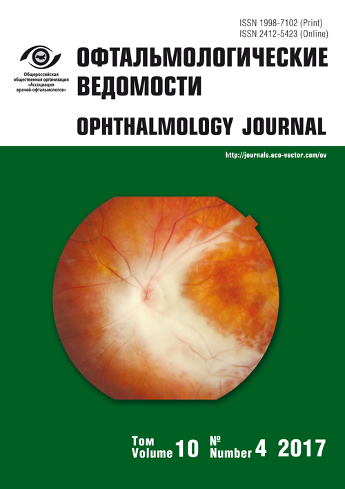
A 29-year-old female patient presented with complaints of decreased visual acuity and object distortion and was admitted to the Ophthalmology Clinic of the Pavlov First Saint Petersburg State Medical University (Saint-Petersburg, Russia) in March 2017. She first noticed these symptoms at the beginning of 2017. Upon examination, her best corrected visual acuity value was 0.5 in the right eye and 1.0 in the left eye. Her intraocular pressure (measured using the Icare tonometer TAOli, Finland) was 11 and 10 mm Hg in the right and left eyes, respectively. Biomicroophthalmoscopy revealed severe vascular tortuosity in the right eye and a large number of thick and twisted vessels in the area of the optic disc and inferior macula. The biomicroophthalmoscopy of the left eye showed no pathological changes. Some vessels were white and sclerosed (Figure 1).

Fig. 1. Fundus photo: a lot of enlarged convoluted blood vessels resembling tangled worms
The patient underwent automated perimetry, optical coherence tomography, fluorescein angiography, and indocyanine green angiography. The automated perimetry revealed blind spot enlargement and absolute scotomas in the inferior part of the central visual field. Optical coherence tomography was used to obtain multiple cross-sectional images of the vessels in the inner layers of the retinal neuroepithelium, to demonstrate the areas of destruction in the retinal pigment epithelium, and an increase in macular volume (Figure 2). Fluorescein angiography of the macular area showed pathologically dilated, tortuous retinal vessels with extremely poor or absent blood circulation (Figure 3a). It also revealed arteriovenous anastomoses and an area of capillary nonperfusion in the upper outer quadrant (Figure 3c). Slight dye leakage in the macular area was detected during the late phase (Figure 3b). Indocyanine green angiography showed no pathological changes in the choroidal blood flow (Figure 4). The patient was referred for brain and orbital MRA, which revealed no pathological changes. No AVMs were detected in the skin. Based on the examination results, we diagnosed the patient with an isolated ocular form of the Wyburn–Mason syndrome. The patient underwent retinal laser photocoagulation in the ischemic area and is currently followed up by a neurosurgeon and an ophthalmologist.

Fig. 2. Optical coherence tomography: increased neuroepithelium thickness in the vascular loops area, vessels at the neuroepithelium inner layers level (white arrows), areas of the retinal pigment epithelium destruction

Fig. 3. Fluorescein angiography: a – early venous phase (28 seconds); b – recirculation phase (12 minutes); c – capillary nonperfusion area in the late phase at the far periphery in the upper-outer quadrant. Description in the text

Fig. 4. Indocyanine green angiography: a – early phase (38 seconds); b – late phase (12 minutes)
About the authors
Yuriy S. Astakhov
Academician I.P. Pavlov First St. Petersburg State Medical University
Author for correspondence.
Email: astakhov73@mail.ru
MD, PhD, DMedSc, professor
Russian Federation, Saint PetersburgSvetlana G. Belekhova
Academician I.P. Pavlov First St. Petersburg State Medical University
Email: beleksv@yandex.ru
MD, assistant
Russian Federation, Saint PetersburgAida A. Shakhnazarova
Diagnostic center № 7 (ophthalmological) for adults and children
Email: beleksv@yandex.ru
MD, PhD
Russian Federation, Saint PetersburgAndranik Yu. Ovnanyan
Academician I.P. Pavlov First St. Petersburg State Medical University
Email: ovnanyan@yandex.ru
MD, assistant. Ophthalmology Department
Russian Federation, Saint PetersburgReferences
- Archer DB, Deutman A, Ernest JT, Krill AE. Arteriovenous communications of the retina. Am J Ophthalmol. 1973;75:224-41. doi: 10.1016/0002-9394(73)91018-0.
- Augsburger JJ, Goldberg RE, Shields JA, et al. Changing appearance of retinal arteriovenous malformation. Albrecht Von Graefes. Arch Klin Exp Ophthalmol. 1980;215:65-70. doi: 10.1007/BF00413398.
- Bhojwani D, Vachhrajani M, Vasavada A. Wyburn Mason Syndrome: A Rare Phacomatosis. Ophthalmology. 2016Aug; 123(8):1787. doi: 10.1016/j.ophtha.2016.04.053.
- Bonnet P, Dechaume J, Blanc E. L’anévrysme cirsoïde de la rétine (anévrysme racémeux). Ses relations avec l’anévrysme cirsoïde de la face et avec l’anévrysme cirsoïde du cerveau. Journal de médecine de Lyon. 1937;18:165-78.
- Brea GB. Angioma encefalo-oculo-faciale (Sindrome di Bonnet, Dechaume e Blanc). Rivista Neurolog. 1963;33:455-74.
- Brock S, Dyke CG. Venous and arteriovenous angiomas of the brain. A clinical and roentgenographic study of eight cases (case 3). Bull Neurol Inst New York. 1932;2:257-65.
- Brodsky MC, Hoyt WF. Spontaneous involution of retinal and intracranial arteriovenous malformation in Bonnet-Dechaume-Blanc syndrome. Br J Ophthalmol. 2002;86:360-1. doi: 10.1136/bjo.86.3.360.
- Chowaniec MJ, Suh DW, Boldt HC, et al. Anomalous optical coherence tomography findings in Wyburn-Mason syndrome and isolated retinal arteriovenous malformation. J AAPOS. 2015Apr;19(2):175-7. doi: 10.1016/j.jaapos.2014.09.019.
- Dayani PN, Sadun AA. A case report of Wyburn-Mason syndrome and review of the literature. Neuroradiology. 2007;49:445-456. doi: 10.1007/s00234-006-0205-x.
- De Keizer RJW, van Dalen JTW. Wyburn-Mason syndrome. Subcutaneous angioma extirpation after preliminary embolisation. Doc Ophthalmol. 1981;50:263-73. doi: 10.1007/BF00158008.
- Dekking HM. Arteriovenous aneurysm of the retina with spontaneous regression. Ophthalmologica. 1955;130:113-5. doi: 10.1159/000302653.
- Effron L, Zakov ZN, Tomsak RL. Neovascular glaucoma as a complication of the Wyburn-Mason syndrome. J Clin Neuroophthalmol. 1985;5:95-98.
- Fujita H, Nakano K, Kumon Y, et al. A case of 1958 Wyburn-Mason syndrome. Rinsho Shinkeigaku. 1989;29:1039-44.
- Glees M. Arteriovenoses Aneurysma des Augenhintergrundes und der gleichseitigen Großhirnhemisphare. Klin Monatsbl Augenheilkd. 1954;124:457-60.
- Krug EF, Samuels B. Venous angioma of the retina, optic nerve, chiasm and brain. A case report with postmortem observations. Arch Ophthalmol. 1932;8:871-9. doi: 10.1001/archopht.1932.00820190091010.
- Maeda H, Fujieda M, Morita H, Kurashige T. Wyburn-Mason syndrome – a case report. No To Hattatsu. 1992;24:65-9.
- Magnus H. Aneurysma arterioso-venosum retinale. Virchows Arch Pathol Anat. 1874;60:38-45. doi: 10.1007/BF01938766.
- Mansour AM, Wells CG, Jampol LM, Kalina RE. Ocular complications of arteriovenous communications of the retina. Arch Ophthalmol. 1989;107:232-236. doi: 10.1001/archopht.1989.01070010238029.
- Muthukumar N, Prabakara Sundaralingam M. Retinocephalic vascular malformation: case report. Br J Neurosurg. 1998;12: 458-60. doi: 10.1080/02688699844718.
- Odom GL. Ophthalmic involvement in neurological vascular lesions, case 14. In: Neuro-Ophthalmology Symposium. Ed by J.L. Smith. University of Miami: Springfield IL, Charles C Thomas Publishers; 1964; P. 244-7.
- Onder HI, Alisan S, Tunc M. Serous retinal detachment and cystoid macular edema in a patient with Wyburn-Mason syndrome. Semin Ophthalmol. 2015 Mar;30(2):154-6. doi: 10.3109/08820538.2013.835832.
- Patel KH, Kalevar A, Mcdonald HR, Johnson RN. Retinal arteriovenous malformation. Retin Cases Brief Rep. 2017Winter;11 Suppl1: S11-S13. doi: 10.1097/ICB.0000000000000384.
- Rao P, Thomas BJ, Yonekawa Y, et al. Peripheral Retinal Ischemia, Neovascularization, and Choroidal Infarction in Wyburn-Mason Syndrome. JAMA Ophthalmol. 2015Jul;133(7):852-4. doi: 10.1001/jamaophthalmol.2015.0716.
- Reck SD, Zacks DN, Eibschitz-Tsimhoni M. Retinal and intracranial arteriovenous malformations: Wyburn-Mason syndrome. J Neuroophthalmol. 2005;25:205-8. doi: 10.1097/01.wno.0000177301.82963.9f.
- Sahoo IM, Sahoo RN, Das BS. Arteriovenous malformation of brain and eye. Indian J Ophthalmol. 1982;30:155-6.
- Schmidt D, Pache M, Schumacher M. The congenital unilateral retinocephalic vascular malformation syndrome (Bonnet-Dechaume-Blanc syndrome or Wyburn-Mason syndrome): review of the literature. Surv Ophthalmol. 2008;53:227-249. doi: 10.1016/j.survophthal.2007.10.001.
- Selhorst JB. Phacomatoses. In: Walsh and Hoyt’s Clinical Neuro-Ophthalmology. Ed by N.R. Miller, N.J. Newman. Baltimore: Williams and Wilkins Co; 1998. Ed 5, vol 2, p. 2725-9, chapter 49.
- Shah GK, Shields JA, Lanning RC. Branch retinal vein obstruction secondary to retinal arteriovenous communication. Am J Ophthalmol. 1998;126:446-448 doi: 10.1016/S0002-9394(98)00103-2.
- Théron J, Newton TH, Hoyt WF. Unilateral retinocephalic vascular malformations. Neuroradiology. 1974;7:185-96. doi: 10.1007/BF00342696.
- Wyburn-Mason R. Arteriovenous aneurysm of midbrain and retina, facial naevi and mental changes. Brain. 1943;66:163-203. doi: 10.1093/brain/66.3.163.
Supplementary files

