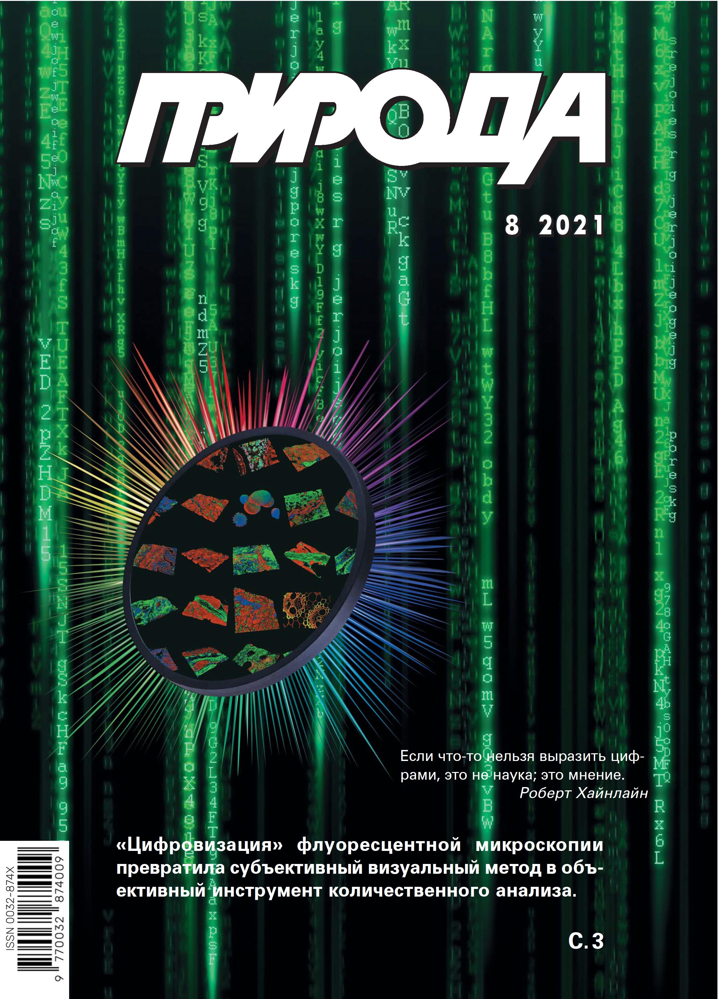
Digital Fluorescence Microscopy: New Analytical Tool for Studying Microorganisms
- Авторлар: Puchkov E.O1,2
-
Мекемелер:
- Skryabin Institute of Biochemistry and Physiology of Microorganisms, Pushchino Center for Biological Research, RAS
- Pushchino State Institute of Natural Science
- Шығарылым: № 8 (2021)
- Беттер: 3-15
- Бөлім: Articles
- URL: https://journals.eco-vector.com/0032-874X/article/view/628015
- DOI: https://doi.org/10.7868/S0032874X21080019
- ID: 628015
Дәйексөз келтіру
Аннотация
Over the last twenty years, fluorescence microscopy has transformed from a subjective visual method to an objective analytical approach, digital fluorescence microscopy (DFM). The real reasons for this are innovations related to modernization of microscopes, computer processing and analysis of digital images, as well as the usage of new fluorophores and methods for their introduction into the cells of microorganisms. DFM has provided fundamentally new possibilities for the study of microorganisms, which allows obtaining unique data on the localization and dynamics of intracellular processes within the study of single cells of microorganisms at the subcellular level.
Авторлар туралы
E. Puchkov
Skryabin Institute of Biochemistry and Physiology of Microorganisms, Pushchino Center for Biological Research, RAS; Pushchino State Institute of Natural Science
Email: puchkov@ibpm.pushchino.ru
Pushchino, Russia; Pushchino, Russia
Әдебиет тізімі
- Lakowicz J.R. Principles of Fluorescence Spectroscopy. Berlin, 2006.
- Puchkov E. Analytical Techniques for Single-Cell Studies in Microbiology. Handbook of Single Cell Technologies. T.Santra, F.G.Tseng (eds.). Singapore, 2021; 1–32. doi: 10.1007/978-981-10-4857-9_17-3.
- Sanderson M.J., Smith I., Parker I. et al. Fluorescence microscopy. Cold Spring Harb Protoc. 2014; 10: pdb.top071795. doi: 10.1101/pdb.top071795.
- Sbalzarini I.F. Seeing is believing, quantifying is convincing, computational image analysis in biology. Adv. Anat. Embryol. Cell Biol. 2016; 219: 1–39. doi: 10.1007/978-3-319-28549-8_1.
- Nketia T.A., Sailem H., Rohde G. et al. Analysis of live cell images. Methods, tools and opportunities. Method. 2017; 115: 65–79. doi: 10.1016/j.ymeth.2017.02.007.
- Wallace C.T., Jessup M., Bernas T. et al. Basics of digital microscopy. Curr. Protoc. Cytom. 2018; 83: 12.2.1–12.2.14. doi: 10.1002/cpcy.31.
- Puchkov E. Quantitative Optical Microscopy in Microbiology. An Introduction to Microorganisms. Q-S.Wu, Y-N.Zou, F.Zhang, B.Shu (eds). N.Y., 2021; Chapter 1: 1–31.
- The Molecular Probes Handbook. A Guide to Fluorescent Probes and Labeling Technologies. I.Johnson, M.Spence (eds.). Moscow: Life Technologies, 2010.
- Tsien R.Y. The green fluorescent protein. Annu. Rev. Biochem. 1998; 67: 509–544. doi: 10.1146/annurev.biochem.67.1.509.
- Remington S.J. Green fluorescent protein: a perspective. Protein Sci. 2011; 20(9): 1509–1519. doi: 10.1002/pro.684.
- Tsien R.Y. Building and breeding molecules to spy on cells and tumors. FEBS Lett. 2005; 579(4): 927–932. doi: 10.1016/j.febslet.2004.11.025.
- Snapp E. Design and use of fluorescent fusion proteins in cell biology. Curr. Protoc. Cell Biol. 2005; Chapter 21: 21.4.1–21.4.13. doi: 10.1002/0471143030.cb2104s27.
- Campbell R.E. Fluorescent proteins. Scholarpedia. 2008; 3(7): 5410. doi: 10.4249/scholarpedia.5410.
- Keppler A., Gendreizig S., Gronemeyer T. et al. A general method for the covalent labeling of fusion proteins with small molecules in vivo. Nat Biotechnol. 2003; 21: 86–89. doi: 10.1038/nbt765.
- Juillerat A., Gronemeyer T., Keppler A. et al. Directed evolution of O6-alkylguanine-DNA alkyltransferase for efficient labeling of fusion proteins with small molecules in vivo. Chem. Biol. 2003; 10(4): 313–317. doi: 10.1016/S1074-5521(03)00068-1.
- Gautier A., Juillerat A., Heinis C. et al. An engineered protein tag for multiprotein labeling in living cells. Chem. Biol. 2008; 15:128–136. doi: 10.1016/j.chembiol.2008.01.007.
- Los G.V., Encell L.P., McDougall M.G. et al. HaloTag, a novel protein labeling technology for cell imaging and protein analysis. ACS Chem. Biol. 2008; 3(6): 373–382. doi: 10.1021/cb800025k.
- Hinner M.J., Johnsson K. How to obtain labeled proteins and what to do with them. Curr. Opin. Biotechnol. 2010; 21(6): 766–776. doi: 10.1016/j.copbio.2010.09.011.
- Chozinski T.J., Gagnon L.A., Vaughan J.C. Twinkle, twinkle little star, photoswitchable fluorophores for super-resolution imaging. FEBS Lett. 2014; 588(19): 3603–3612. doi: 10.1016/j.febslet.2014.06.043.
- Minoshima M., Kikuchi K. Photostable and photoswitching fluorescent dyes for super-resolution imaging. J. Biol. Inorg. Chem. 2017; 22(5): 639–652. doi: 10.1007/s00775-016-1435-y.
- Hell S. Far-Field Optical nanoscopy. Science. 2007; 316(5828): 1153–1158. doi: 10.1126/science.1137395.
- Betzig E., Patterson G.H., Sougrat R. et al. Imaging intracellular fluorescent proteins at nanometer resolution. Science. 2006; 313(5793): 1642–1645. doi: 10.1126/science.1127344.
- Hess S.T., Girirajan T.P., Mason M.D. Ultra-high resolution imaging by fluorescence photoactivation localization microscopy. Biophys. J. 2006; 91(11): 4258–4272. doi: 10.1529/biophysj.106.091116.
- Rust M.J., Bates M., Zhuang X. Sub-diffraction-limit imaging by stochastic optical reconstruction microscopy (STORM). Nat. Methods. 2006; 3(10): 793–795. doi: 10.1038/nmeth929.
- Bates M., Huang B., Dempsey G.T. et al. Multicolor super-resolution imaging with photoswitchable fluorescent probes. Science. 2007; 317(5845): 1749–1753. doi: 10.1126/science.1146598.
- Heilemann M., Dedecker P., Hofkens J. et al. Photoswitches: key molecules for subdiffraction resolution fluorescence imaging and molecular quantification. Laser Photon. Rev. 2009; 3(1–2): 180–202. doi: 10.1002/lpor.200810043.
- Klar T.A., Jakobs S., Dyba M. et al. Fluorescence microscopy with diffraction resolution barrier broken by stimulated emission. Proc. Natl. Acad. Sci. USA. 2000; 97(15): 8206–8210. doi: 10.1073/pnas.97.15.8206.
- Gustafsson M.G.L. Nonlinear structured-illumination microscopy: wide-field fluorescence imaging with theoretically unlimited resolution. Proc. Natl. Acad. Sci. USA. 2005; 102(37): 13081–13086. doi: 10.1073/pnas.0406877102.
- Пучков Е.О. Внутриклеточная вязкость: методы измерения и роль в метаболизме. Биологические мембраны. 2014; 31(1): 3–13. doi: 10.7868/80233475513050149.
- Puchkov E. Microfluorimetry of Single Yeast Cells by Fluorescence Microscopy Combined with Digital Photography and Computer Image Analysis. Advances in Medicine and Biology. L.V.Berhardt (ed.). N.Y., 2016; 98: 69–90. doi: 10.1134/S0026365619010130.
- Ohtani M., Saka A., Sano F. et al. Development of image processing program for yeast cell morphology. J. Bioinform. Computat. Biol. 2004; 1(4): 695–709. doi: 10.1142/s0219720004000363.
- Negishi T., Nogami S., Ohya Y. Multidimensional quantification of subcellular morphology of Saccharomyces cerevisiae using CalMorph, the high-throughput image-processing program. J. Biotechnol. 2009; 141(3–4): 109–117. doi: 10.1016/j.jbiotec.2009.03.014.
- Nogami S., Ohya Y., Yvert G. Genetic complexity and quantitative trait loci mapping of yeast morphological traits. PLoS Genet. 2007; 3(2): e31. doi: 10.1371/journal.pgen.0030031.
- Gebre A.A., Okada H., Kim C. et al. Profiling of the effects of antifungal agents on yeast cells based on morphometric analysis. FEMS Yeast Res. 2015; 15(5): fov040. doi: 10.1093/femsyr/fov040.
- Bjerling P., Olsson I., Meng X. Quantitative live cell fluorescence-microscopy analysis of fission yeast. J. Vi. Exp. 2012; 59: e3454. doi: 10.3791/3454.
- Akamatsu M., Lin Y., Bewersdorf J. et al. Analysis of interphase node proteins in fission yeast by quantitative and superresolution fluorescence microscopy. Mol. Biol. Cell. 2017; 28(23): 3203–3214. doi: 10.1091/mbc.E16-07-0522.
- Arasada R., Sayyad W.A., Berro J. et al. High-speed superresolution imaging of the proteins in fission yeast clathrin-mediated endocytic actin patches. Mol. Biol. Cell. 2018; 29(3): 295–303. doi: 10.1091/mbc.E17-06-0415.
- Bestul A.J., Yu Z., Unruh J.R., Jaspersen S.L. Molecular model of fission yeast centrosome assembly determined by superresolution imaging. J. Cell Biol. 2017; 216(8): 2409–2424. doi: 10.1083/jcb.201701041.
- Wollman A., Hedlund E.G., Shashkova S. et al. Towards mapping the 3D genome through high speed single-molecule tracking of functional transcription factors in single living cells. Methods. 2020; 170: 82–89. doi: 10.1016/j.ymeth.2019.06.021.
- Elowitz M.B., Surette M.G., Wolf P.E. et al. Photoactivation turns green fluorescent protein red. Curr Biol. 1997; 7(10): 809–812. doi: 10.1016/s0960-9822(06)00342-3.
- Uphoff S., Reyes-Lamothe R., de Leon F.G. et al. Single-molecule DNA repair in live bacteria. PNAS. 2013; 110(20): 8063–8068. doi: 10.1073/pnas.1301804110.
- Virant D., Turkowyd B., Balinovic A. et al. Combining primed photoconversion and UV-photoactivation for aberration-free, live-cell compliant multi-color single-molecule localization microscopy imaging. Int. J. Mol. Sci. 2017; 18(7): 1524. doi: 10.3390/ijms18071524.
- Yao Z., Carballido-Lуpez R. Fluorescence imaging for bacterial cell biology, from localization to dynamics, from ensembles to single molecules. Annu. Rev. Microbiol. 2014; 68: 459–476. doi: 10.1146/annurev-micro-091213-113034.
- Stracy M., Kapanidis A.N. Single-molecule and super-resolution imaging of transcription in living bacteria. Methods. 2017; 120: 103–114. doi: 10.1016/j.ymeth.2017.04.001.
- Gahlmann A., Moerner W.E. Exploring bacterial cell biology with single-molecule tracking and super-resolution imaging. Nat. Rev. Microbiol. 2014; 12(1): 9–22. doi: 10.1038/nrmicro3154.
- Uphoff S. Super-resolution microscopy and tracking of DNA-binding proteins in bacterial cells. Methods Mol. Biol. 2016; 1431: 221–234. doi: 10.1007/978-1-4939-3631-1_16.
- Li Y., Schroeder J.W., Simmons L.A. et al. Visualizing bacterial DNA replication and repair with molecular resolution. Curr Opin Microbiol. 2018; 43: 38–45. doi: 10.1016/j.mib.2017.11.009.
- Schneider J.P., Basler M. Shedding light on biology of bacterial cells. Phil. Trans. R. Soc B. 2016; 371(1707): 20150499. doi: 10.1098/rstb.2015.0499.
- Kentner D., Sourjik V. Use of fluorescence microscopy to study intracellular signaling in bacteria. Annu. Rev. Microbiol. 2010; 64: 373–390. doi: 10.1146/annurev.micro.112408.134205.
- Choi H., Rangarajan N., Weisshaar J.C. Lights, camera, action! Antimicrobial peptide mechanisms imaged in space and time. Trends Microbiol. 2016; 24(2): 111–122. doi: 10.1016/j.tim.2015.11.004.
- Haas B.L., Matson J.S., DiRita V.J. et al. Imaging live cells at the nanometer-scale with single-molecule microscopy, obstacles and achievements in experiment optimization for microbiology. Molecules. 2014; 19(8): 12116–12149. doi: 10.3390/molecules190812116.
- Endesfelder U. From single bacterial cell imaging towards in vivo single-molecule biochemistry studies. Essays Biochem. 2019; 63(2): 187–196. doi: 10.1042/EBC20190002.
Қосымша файлдар


