


")


Цифровая флуоресцентная микроскопия: новый аналитический инструмент для изучения микроорганизмов
- Авторы: Пучков Е.О1,2
-
Учреждения:
- Институт биохимии и физиологии микроорганизмов имени Г.К.Скрябина РАН Федерального исследовательского центра «Пущинский научный центр биологических исследований РАН»
- Пущинский государственный естественно-научный институт
- Выпуск: № 8 (2021)
- Страницы: 3-15
- Раздел: Статьи
- URL: https://journals.eco-vector.com/0032-874X/article/view/628015
- DOI: https://doi.org/10.7868/S0032874X21080019
- ID: 628015
Цитировать
Полный текст

Аннотация
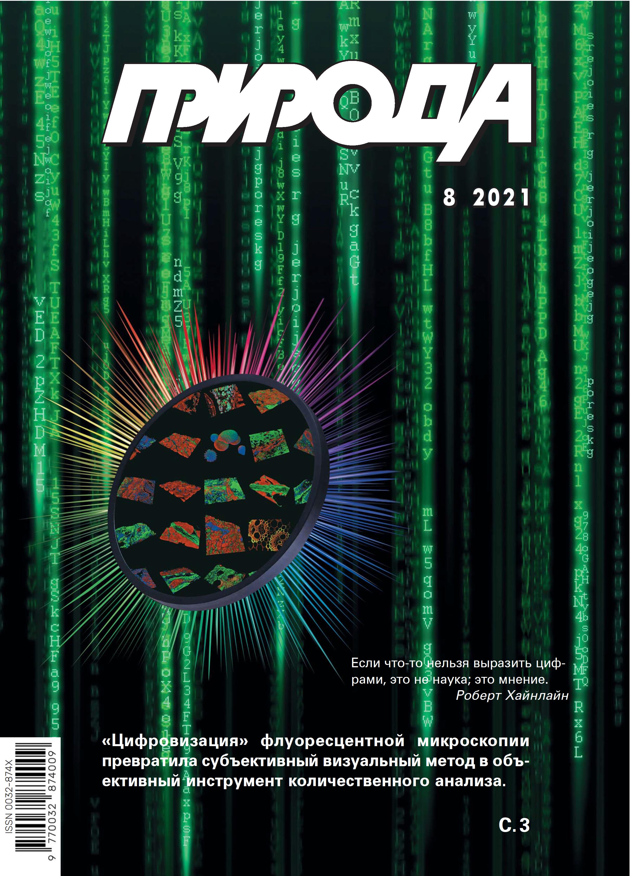
За последние двадцать лет флуоресцентная микроскопия трансформировалась из субъективного визуального метода в объективный аналитический подход — цифровую флуоресцентную микроскопию (ЦФМ). Произошло это благодаря совокупности инноваций, связанных с модернизацией микроскопов, использованием компьютерной обработки и анализа цифровых изображений, а также применением новых флуорофоров и методов их внедрения в клетки микроорганизмов. ЦФМ открыла принципиально новые возможности для изучения микроорганизмов, которые позволяют получать уникальную информацию о локализации и динамике внутриклеточных процессов при изучении единичных клеток микроорганизмов на субклеточном уровне.
Об авторах
Е. О Пучков
Институт биохимии и физиологии микроорганизмов имени Г.К.Скрябина РАН Федерального исследовательского центра «Пущинский научный центр биологических исследований РАН»; Пущинский государственный естественно-научный институт
Email: puchkov@ibpm.pushchino.ru
Пущино, Россия; Пущино, Россия
Список литературы
- Lakowicz J.R. Principles of Fluorescence Spectroscopy. Berlin, 2006.
- Puchkov E. Analytical Techniques for Single-Cell Studies in Microbiology. Handbook of Single Cell Technologies. T.Santra, F.G.Tseng (eds.). Singapore, 2021; 1–32. doi: 10.1007/978-981-10-4857-9_17-3.
- Sanderson M.J., Smith I., Parker I. et al. Fluorescence microscopy. Cold Spring Harb Protoc. 2014; 10: pdb.top071795. doi: 10.1101/pdb.top071795.
- Sbalzarini I.F. Seeing is believing, quantifying is convincing, computational image analysis in biology. Adv. Anat. Embryol. Cell Biol. 2016; 219: 1–39. doi: 10.1007/978-3-319-28549-8_1.
- Nketia T.A., Sailem H., Rohde G. et al. Analysis of live cell images. Methods, tools and opportunities. Method. 2017; 115: 65–79. doi: 10.1016/j.ymeth.2017.02.007.
- Wallace C.T., Jessup M., Bernas T. et al. Basics of digital microscopy. Curr. Protoc. Cytom. 2018; 83: 12.2.1–12.2.14. doi: 10.1002/cpcy.31.
- Puchkov E. Quantitative Optical Microscopy in Microbiology. An Introduction to Microorganisms. Q-S.Wu, Y-N.Zou, F.Zhang, B.Shu (eds). N.Y., 2021; Chapter 1: 1–31.
- The Molecular Probes Handbook. A Guide to Fluorescent Probes and Labeling Technologies. I.Johnson, M.Spence (eds.). Moscow: Life Technologies, 2010.
- Tsien R.Y. The green fluorescent protein. Annu. Rev. Biochem. 1998; 67: 509–544. doi: 10.1146/annurev.biochem.67.1.509.
- Remington S.J. Green fluorescent protein: a perspective. Protein Sci. 2011; 20(9): 1509–1519. doi: 10.1002/pro.684.
- Tsien R.Y. Building and breeding molecules to spy on cells and tumors. FEBS Lett. 2005; 579(4): 927–932. doi: 10.1016/j.febslet.2004.11.025.
- Snapp E. Design and use of fluorescent fusion proteins in cell biology. Curr. Protoc. Cell Biol. 2005; Chapter 21: 21.4.1–21.4.13. doi: 10.1002/0471143030.cb2104s27.
- Campbell R.E. Fluorescent proteins. Scholarpedia. 2008; 3(7): 5410. doi: 10.4249/scholarpedia.5410.
- Keppler A., Gendreizig S., Gronemeyer T. et al. A general method for the covalent labeling of fusion proteins with small molecules in vivo. Nat Biotechnol. 2003; 21: 86–89. doi: 10.1038/nbt765.
- Juillerat A., Gronemeyer T., Keppler A. et al. Directed evolution of O6-alkylguanine-DNA alkyltransferase for efficient labeling of fusion proteins with small molecules in vivo. Chem. Biol. 2003; 10(4): 313–317. doi: 10.1016/S1074-5521(03)00068-1.
- Gautier A., Juillerat A., Heinis C. et al. An engineered protein tag for multiprotein labeling in living cells. Chem. Biol. 2008; 15:128–136. doi: 10.1016/j.chembiol.2008.01.007.
- Los G.V., Encell L.P., McDougall M.G. et al. HaloTag, a novel protein labeling technology for cell imaging and protein analysis. ACS Chem. Biol. 2008; 3(6): 373–382. doi: 10.1021/cb800025k.
- Hinner M.J., Johnsson K. How to obtain labeled proteins and what to do with them. Curr. Opin. Biotechnol. 2010; 21(6): 766–776. doi: 10.1016/j.copbio.2010.09.011.
- Chozinski T.J., Gagnon L.A., Vaughan J.C. Twinkle, twinkle little star, photoswitchable fluorophores for super-resolution imaging. FEBS Lett. 2014; 588(19): 3603–3612. doi: 10.1016/j.febslet.2014.06.043.
- Minoshima M., Kikuchi K. Photostable and photoswitching fluorescent dyes for super-resolution imaging. J. Biol. Inorg. Chem. 2017; 22(5): 639–652. doi: 10.1007/s00775-016-1435-y.
- Hell S. Far-Field Optical nanoscopy. Science. 2007; 316(5828): 1153–1158. doi: 10.1126/science.1137395.
- Betzig E., Patterson G.H., Sougrat R. et al. Imaging intracellular fluorescent proteins at nanometer resolution. Science. 2006; 313(5793): 1642–1645. doi: 10.1126/science.1127344.
- Hess S.T., Girirajan T.P., Mason M.D. Ultra-high resolution imaging by fluorescence photoactivation localization microscopy. Biophys. J. 2006; 91(11): 4258–4272. doi: 10.1529/biophysj.106.091116.
- Rust M.J., Bates M., Zhuang X. Sub-diffraction-limit imaging by stochastic optical reconstruction microscopy (STORM). Nat. Methods. 2006; 3(10): 793–795. doi: 10.1038/nmeth929.
- Bates M., Huang B., Dempsey G.T. et al. Multicolor super-resolution imaging with photoswitchable fluorescent probes. Science. 2007; 317(5845): 1749–1753. doi: 10.1126/science.1146598.
- Heilemann M., Dedecker P., Hofkens J. et al. Photoswitches: key molecules for subdiffraction resolution fluorescence imaging and molecular quantification. Laser Photon. Rev. 2009; 3(1–2): 180–202. doi: 10.1002/lpor.200810043.
- Klar T.A., Jakobs S., Dyba M. et al. Fluorescence microscopy with diffraction resolution barrier broken by stimulated emission. Proc. Natl. Acad. Sci. USA. 2000; 97(15): 8206–8210. doi: 10.1073/pnas.97.15.8206.
- Gustafsson M.G.L. Nonlinear structured-illumination microscopy: wide-field fluorescence imaging with theoretically unlimited resolution. Proc. Natl. Acad. Sci. USA. 2005; 102(37): 13081–13086. doi: 10.1073/pnas.0406877102.
- Пучков Е.О. Внутриклеточная вязкость: методы измерения и роль в метаболизме. Биологические мембраны. 2014; 31(1): 3–13. doi: 10.7868/80233475513050149.
- Puchkov E. Microfluorimetry of Single Yeast Cells by Fluorescence Microscopy Combined with Digital Photography and Computer Image Analysis. Advances in Medicine and Biology. L.V.Berhardt (ed.). N.Y., 2016; 98: 69–90. doi: 10.1134/S0026365619010130.
- Ohtani M., Saka A., Sano F. et al. Development of image processing program for yeast cell morphology. J. Bioinform. Computat. Biol. 2004; 1(4): 695–709. doi: 10.1142/s0219720004000363.
- Negishi T., Nogami S., Ohya Y. Multidimensional quantification of subcellular morphology of Saccharomyces cerevisiae using CalMorph, the high-throughput image-processing program. J. Biotechnol. 2009; 141(3–4): 109–117. doi: 10.1016/j.jbiotec.2009.03.014.
- Nogami S., Ohya Y., Yvert G. Genetic complexity and quantitative trait loci mapping of yeast morphological traits. PLoS Genet. 2007; 3(2): e31. doi: 10.1371/journal.pgen.0030031.
- Gebre A.A., Okada H., Kim C. et al. Profiling of the effects of antifungal agents on yeast cells based on morphometric analysis. FEMS Yeast Res. 2015; 15(5): fov040. doi: 10.1093/femsyr/fov040.
- Bjerling P., Olsson I., Meng X. Quantitative live cell fluorescence-microscopy analysis of fission yeast. J. Vi. Exp. 2012; 59: e3454. doi: 10.3791/3454.
- Akamatsu M., Lin Y., Bewersdorf J. et al. Analysis of interphase node proteins in fission yeast by quantitative and superresolution fluorescence microscopy. Mol. Biol. Cell. 2017; 28(23): 3203–3214. doi: 10.1091/mbc.E16-07-0522.
- Arasada R., Sayyad W.A., Berro J. et al. High-speed superresolution imaging of the proteins in fission yeast clathrin-mediated endocytic actin patches. Mol. Biol. Cell. 2018; 29(3): 295–303. doi: 10.1091/mbc.E17-06-0415.
- Bestul A.J., Yu Z., Unruh J.R., Jaspersen S.L. Molecular model of fission yeast centrosome assembly determined by superresolution imaging. J. Cell Biol. 2017; 216(8): 2409–2424. doi: 10.1083/jcb.201701041.
- Wollman A., Hedlund E.G., Shashkova S. et al. Towards mapping the 3D genome through high speed single-molecule tracking of functional transcription factors in single living cells. Methods. 2020; 170: 82–89. doi: 10.1016/j.ymeth.2019.06.021.
- Elowitz M.B., Surette M.G., Wolf P.E. et al. Photoactivation turns green fluorescent protein red. Curr Biol. 1997; 7(10): 809–812. doi: 10.1016/s0960-9822(06)00342-3.
- Uphoff S., Reyes-Lamothe R., de Leon F.G. et al. Single-molecule DNA repair in live bacteria. PNAS. 2013; 110(20): 8063–8068. doi: 10.1073/pnas.1301804110.
- Virant D., Turkowyd B., Balinovic A. et al. Combining primed photoconversion and UV-photoactivation for aberration-free, live-cell compliant multi-color single-molecule localization microscopy imaging. Int. J. Mol. Sci. 2017; 18(7): 1524. doi: 10.3390/ijms18071524.
- Yao Z., Carballido-Lуpez R. Fluorescence imaging for bacterial cell biology, from localization to dynamics, from ensembles to single molecules. Annu. Rev. Microbiol. 2014; 68: 459–476. doi: 10.1146/annurev-micro-091213-113034.
- Stracy M., Kapanidis A.N. Single-molecule and super-resolution imaging of transcription in living bacteria. Methods. 2017; 120: 103–114. doi: 10.1016/j.ymeth.2017.04.001.
- Gahlmann A., Moerner W.E. Exploring bacterial cell biology with single-molecule tracking and super-resolution imaging. Nat. Rev. Microbiol. 2014; 12(1): 9–22. doi: 10.1038/nrmicro3154.
- Uphoff S. Super-resolution microscopy and tracking of DNA-binding proteins in bacterial cells. Methods Mol. Biol. 2016; 1431: 221–234. doi: 10.1007/978-1-4939-3631-1_16.
- Li Y., Schroeder J.W., Simmons L.A. et al. Visualizing bacterial DNA replication and repair with molecular resolution. Curr Opin Microbiol. 2018; 43: 38–45. doi: 10.1016/j.mib.2017.11.009.
- Schneider J.P., Basler M. Shedding light on biology of bacterial cells. Phil. Trans. R. Soc B. 2016; 371(1707): 20150499. doi: 10.1098/rstb.2015.0499.
- Kentner D., Sourjik V. Use of fluorescence microscopy to study intracellular signaling in bacteria. Annu. Rev. Microbiol. 2010; 64: 373–390. doi: 10.1146/annurev.micro.112408.134205.
- Choi H., Rangarajan N., Weisshaar J.C. Lights, camera, action! Antimicrobial peptide mechanisms imaged in space and time. Trends Microbiol. 2016; 24(2): 111–122. doi: 10.1016/j.tim.2015.11.004.
- Haas B.L., Matson J.S., DiRita V.J. et al. Imaging live cells at the nanometer-scale with single-molecule microscopy, obstacles and achievements in experiment optimization for microbiology. Molecules. 2014; 19(8): 12116–12149. doi: 10.3390/molecules190812116.
- Endesfelder U. From single bacterial cell imaging towards in vivo single-molecule biochemistry studies. Essays Biochem. 2019; 63(2): 187–196. doi: 10.1042/EBC20190002.
Дополнительные файлы
