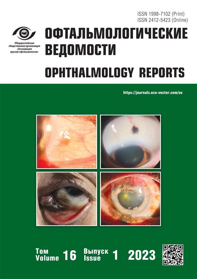
The use of optical coherence tomography angiography in differential diagnosis of conjunctival melanocytic tumors
- Authors: Kiseleva T.N.1, Saakyan S.V.1,2, Makukhina V.V.1, Lugovkina K.V.1, Milash S.V.1, Musova N.F.1, Zharov A.A.1
-
Affiliations:
- Helmholz National Medical Research Center of Eye Diseases
- A.I. Yevdokimov Moscow State University of Medicine and Dentistry
- Issue: Vol 16, No 1 (2023)
- Pages: 27-37
- Section: Original study articles
- Submitted: 01.02.2023
- Accepted: 03.03.2023
- Published: 09.04.2023
- URL: https://journals.eco-vector.com/ov/article/view/173174
- DOI: https://doi.org/10.17816/OV173174
- ID: 173174
Cite item
Abstract
BACKGROUND: Optical coherence tomography angiography (OCTA) is a noninvasive method of eye microcirculation evaluation. Few reports are published on the use of OCTA for anterior segment (AS) vessels analysis in healthy eyes and in conjunctival tumors, and their vascular characteristics are still not thoroughly investigated. These questions are of importance, as it is known that tumor’s vasculature is indicative of the patient’s vital prognosis.
AIM: The aim of our study was to investigate the potential of AS-OCTA in evaluation of normal conjunctival vessels architecture as well as that in melanocytic neoplasms.
MATERIALS AND METHODS: 20 healthy volunteers (20 eyes) and 20 patients (20 eyes) with conjunctival nevi and melanomas were examined. AS optical coherence tomography (OCT) and AS-OCTA were performed. Scan analysis included qualitative assessment (vessels pattern, lumen, tortuosity) and quantitative assessment [perfusion density (PD, %) index]. Mean (MPD), maximum (MaxPD) and perifocal PD (PPD) were determined.
RESULTS: In normal group, predominantly radial pattern of the vessels was revealed, their caliber remaining the same along their entire length; larger vessels were more often discovered in deep conjunctival layers. The lowest PD value (29.9%) was registered in the inferior conjunctival segment, and the highest (36.7%) — in the nasal one. In the conjunctival tumors’ area tortuosity of the vessels, uneven vessels’ caliber along their length, and increase in the PD value were observed. Melanomas were characterized by an increase in the “lace-like pattern” and by presence of “confluent pattern” zones; MaxPD value was more than 50%. Significant difference was found between MPD values of normal conjunctiva and MPD values in conjunctival melanoma.
CONCLUSIONS: AS-OCTA is an informative method for the visualization of vessels in normal conjunctiva and in conjunctival tumors. If the tumor’s vessels are unevenly distributed, MaxPD should be measured.
Full Text
About the authors
Tatiana N. Kiseleva
Helmholz National Medical Research Center of Eye Diseases
Email: tkisseleva@yandex.ru
ORCID iD: 0000-0002-9185-6407
SPIN-code: 5824-5991
Scopus Author ID: 7006275699
Dr. Sci. (Med.), professor, head of Ultrasound diagnostic Department
Russian Federation, MoscowSvetlana V. Saakyan
Helmholz National Medical Research Center of Eye Diseases; A.I. Yevdokimov Moscow State University of Medicine and Dentistry
Email: svsaakyan@yandex.ru
ORCID iD: 0000-0001-8591-428X
SPIN-code: 4783-9193
Scopus Author ID: 6602897459
corresponding member of the Russian Academy of Sciences, Dr. Sci. (Med.), professor
Russian Federation, Moscow; MoscowViktoriia V. Makukhina
Helmholz National Medical Research Center of Eye Diseases
Author for correspondence.
Email: makuhvik@mail.ru
ORCID iD: 0000-0002-6238-309X
SPIN-code: 6891-8162
Scopus Author ID: 57203354833
postgraduate student
Russian Federation, MoscowKsenia V. Lugovkina
Helmholz National Medical Research Center of Eye Diseases
Email: ksushalyg@mail.ru
ORCID iD: 0000-0002-3531-3846
SPIN-code: 9919-6167
Scopus Author ID: 57200173937
Cand. Sci. (Med.), senior research associate of Ultrasound diagnostic Department
Russian Federation, MoscowSergey V. Milash
Helmholz National Medical Research Center of Eye Diseases
Email: sergey_milash@yahoo.com
ORCID iD: 0000-0002-3553-9896
SPIN-code: 5224-4319
Scopus Author ID: 55924655900
Cand. Sci. (Med.), research associate of Department of refraction pathology, binocular vision and ophthalmoergonomics
Russian Federation, MoscowNelly F. Musova
Helmholz National Medical Research Center of Eye Diseases
Email: nelly_smile@mail.ru
ORCID iD: 0000-0003-0908-6018
ophthalmologist of the Oncology Department of the outpatient clinic
Russian Federation, MoscowAndrey A. Zharov
Helmholz National Medical Research Center of Eye Diseases
Email: and-zarus@yandex.ru
ORCID iD: 0000-0003-1103-6570
SPIN-code: 7272-3765
Scopus Author ID: 58023722600
research associate of Pathomorphology Department
Russian Federation, MoscowReferences
- Saakyan SV, Tatskov RA, Ivanova OA, et al. Surgical treatment of epibulbar malformations. Ophthalmology in Russia. 2019;16(3): 289–295. (In Russ.) doi: 10.18008/1816-5095-2019-3-289-295
- Shields CL, Alset AE, Boal NS, et al. Conjunctival tumors in 5002 cases. Comparative analysis of benign versus malignant counterparts. The 2016 James D. Allen Lecture. Am J Ophthalmol. 2017;173:106–133. doi: 10.1016/j.ajo.2016.09.034
- Ivanova OA. Klinicheskie osobennosti nevusov konjyunktivy v vozrastnom aspekte i optimizatsiya ikh rannego lecheniya [dissertation]. Moscow, 2011. 107 p. (In Russ.)
- Neroev BB, KiselevoĬ TN, editors. Ultrazvukovye issledovaniya v oftalmologii: Rukovodstvo dlya vrachei. 1-e izd. Moscow: IKAR, 2019. 322 p. (In Russ.)
- Zaharova MA, Kuroedov AV. Optic coherent tomography — technology which became a reality. Russian Journal of Clinical Ophthalmology. 2015;(4):204–211. (In Russ.)
- Konopińska J, Lisowski Ł, Wasiluk E, et al. The effectiveness of ultrasound biomicroscopic and anterior segment optical coherence tomography in the assessment of anterior segment tumors: long-term follow-up. J Ophthalmol. 2020;2020:9053737. doi: 10.1155/2020/9053737
- Skalet AH, Li Y, Lu CD, et al. Optical coherence tomography angiography characteristics of iris melanocytic tumors. Ophthalmology. 2017;124(2):197–204. doi: 10.1016/j.ophtha.2016.10.003
- Amiryan AG, Saakyan SV. Prognostic factors for uveal melanoma. The Russian Annals of Ophthalmology. 2015;(1):9095. (In Russ.) doi: 10.17116/oftalma2015131190-94
- Allegrini D, Montesano G, Pece A. Optical coherence tomography angiography of iris nevus: a case report. Case Rep Ophthalmol. 2016;7(3):172–178. doi: 10.1159/000450572
- Kiseleva TN, Kotelin VI, Losanova OA, Lugovkina KV. Noninvasive methods assessment blood flow in anterior segment and clinical application perspective. Ophthalmology in Russia. 2017;14(4):283–290. (In Russ.) doi: 10.18008/1816-5095-2017-4-283-290
- Lumbroso B, Huang D, Jia Y, et al. Clinical guide to angio-OCT: non invasive, dyeless OCT Angiography. New Delhi: Jaypee Brothers, Medical Publishers, 2014.
- Lee WD, Devarajan K, Chua J, et al. Optical coherence tomography angiography for the anterior segment. Eye Vis (Lond). 2019;6:4. doi: 10.1186/s40662-019-0129-2
- Akagi T, Uji A, Huang AS, et al. Conjunctival and intrascleral vasculatures assessed using anterior segment optical coherence tomo graphy angiography in normal eyes. Am J Ophthalmol. 2018;196:1–9. doi: 10.1016/j.ajo.2018.08.009
- Binotti WW, Mills H, Nosé RM, et al. Anterior segment optical coherence tomography angiography in the assessment of ocular surface lesions. Ocul Surf. 2021;22:86–93. doi: 10.1016/j.jtos.2021.07.009
- Aicher NT, Nagahori K, Inoue M, et al. Vascular density of the anterior segment of the eye determined by optical coherence tomography angiography and slit-lamp photography. Ophthalmic Res. 2020;63(6):572–579. doi: 10.1159/000506953
- Mehta N, Liu K, Alibhai AY, et al. Impact of binarization thresholding and brightness/contrast adjustment methodology on optical coherence tomography angiography image quantification. Am J Ophthalmol. 2019;205:54–65. doi: 10.1016/j.ajo.2019.03.008
- Nampei K, Oie Y, Kiritoshi S, et al. Comparison of ocular surface squamous neoplasia and pterygium using anterior segment optical coherence tomography angiography. Am J Ophthalmol Case Rep. 2020;20:100902. doi: 10.1016/j.ajoc.2020.100902
- Liu Z, Wang H, Jiang H, et al. Quantitative analysis of conjunctival microvasculature imaged using optical coherence tomography angio graphy. Eye Vis (Lond). 2019;6:5. doi: 10.1186/s40662-019-0130-9
- Kiseleva TN, Saakyan SV, Makukhina VV, et al. Use of optical coherence tomography angiography in assessment in conjunctival vascular architecture in health and pathology. The Russian Annals of Ophthalmo logy. 2022;138(6):32–42. (In Russ.) doi: 10.17116/oftalma202213806132
- Ivanova OA, Saakyan SV. An experience of using optical coherence tomography in the complex diagnosis of epibulbar tumors. Russian Ophthalmological Journal. 2011;4(1):77–79. (In Russ.)
- Brouwer NJ, Marinkovic M, Bleeker JC, et al. Anterior segment OCTA of melanocytic lesions of the conjunctiva and iris. Am J Ophthalmol. 2021;222:137–147. doi: 10.1016/j.ajo.2020.09.009
- Spaide RF, Fujimoto JG, Waheed NK, et al. Optical coherence tomography angiography. Prog Retin Eye Res. 2018;64:1–55. doi: 10.1016/j.preteyeres.2017.11.003
- Sampson DM, Dubis AM, Chen FK, et al. Towards standardizing retinal optical coherence tomography angiography: a review. Light Sci Appl. 2022;11(1):63. doi: 10.1038/s41377-022-00740-9
- Iovino C, Peiretti E, Braghiroli M, et al. Imaging of iris vasculature: current limitations and future perspective. Eye (Lond). 2022;36(5):930–940. doi: 10.1038/s41433-021-01809-2
- Foo VHX, Ke M, Tan CQL, et al. Anterior segment optical coherence tomography angiography assessment of corneal vascularisation after combined fine-needle diathermy with subconjunctival ranibizumab: a pilot study. Adv Ther. 2021;38(8):4333–4343. doi: 10.1007/s12325-021-01849-w
Supplementary files


