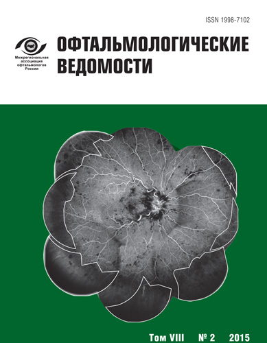
Ranibizumab and retinal photocoagulation in the treatment of ischemic retinal vein occlusion
- Authors: Tultseva S.N.1, Astakhov Y.S.1, Nechiporenko P.A.1, Ovnanyan A.Y.1, Khatina V.A.1
-
Affiliations:
- Pavlov First State Medical University of Saint Petersburg
- Issue: Vol 8, No 2 (2015)
- Pages: 11-27
- Section: Articles
- Submitted: 24.06.2015
- Published: 15.06.2015
- URL: https://journals.eco-vector.com/ov/article/view/372
- DOI: https://doi.org/10.17816/OV2015211-27
- ID: 372
Cite item
Full Text
Abstract
About the authors
Svetlana Nikolaevna Tultseva
Pavlov First State Medical University of Saint Petersburg
Email: tultceva@yandex.ru
MD, professor. Ophthalmology Department
Yury Sergeevich Astakhov
Pavlov First State Medical University of Saint Petersburg
Email: astakhov73@mail.ru
MD, professor. Ophthalmology Department
Pavel Andreevich Nechiporenko
Pavlov First State Medical University of Saint Petersburg
Email: glaz@doctor.com
MD, PhD, assistant professor. Ophthalmology Department
Andranik Yuraevich Ovnanyan
Pavlov First State Medical University of Saint Petersburg
Email: ovnanyan@yandex.ru
ophthalmologist. Ophthalmology Department
Varvara Andreevna Khatina
Pavlov First State Medical University of Saint Petersburg
Email: varvarenka92@mail.ru
medical student. Ophthalmology Department
References
- Кузьмин А. Г., Смирнова О. М., Липатов Д. В., Шестакова М. В. Перспективы лечения диабетической ретинопатии: воздействие на фактор роста эндотелия. Сахарный диабет. 2009; 2: 33-8.
- Тульцева С. Н. Роль воспаления в патогенезе посттромботического макулярного отека. Современные направления медикаментозного лечения. Офтальмологические ведомости. 2012; V (4): 45-51.
- Тульцева С. Н. Значение гипергомоцистеинемии в патогенезе ишемического тромбоза вен сетчатки. Офтальмологические ведомости. 2008; 1 (3): 31-9.
- Тульцева С. Н. Окклюзии вен сетчатки (этиология, патогенез, клиника, диагностика, лечение) / С. Н. Тульцева, Ю. С. Астахов. СПб.: Изд. Н-Л, 2010; 125.
- Тульцева С. Н. Роль наследственных и приобретенных факторов тромбофилии в патогенезе окклюзий вен сетчатки. Автореф. д. м.н., СПб. 2014; 34.
- Baba T., Bikbova G., Kitahashi M. et al. Level of vascular endothelial growth factor 165b in human aqueous humor. Curr Eye Res. 2014; 39 (8): 830-6.
- Boyd S. R., Zachary I., Chakravarthy U. et al. Correlation of increased vascular endothelial growth factor with neovascularization and permeability in ischemic central vein occlusion. Arch Ophthalmol. 2002; 120: 1644-5.
- Boyer D., Heier J., Brown D. M., et al. Vascular endothelial growth factor trap-eye for macular edema secondary to central retinal vein occlusion: six-month results of the phase 3 COPERNICUS study. Ophthalmology. 2012; 119: 1024-32.
- Brown D. M., Campochiaro P. A., Singh R. P. et al. CRUISE Investigators. Ranibizumab for macular edema following central retinal vein occlusion: six-month primary end point results of a phase III study. Ophthalmology. 2010; 117: 1124-33.
- Brown D. M., Wykoff C. C., Wong T. P. et al. Ranibizumab in pre-proliferative (ischemic) central retinal vein occlusion (CRVO): the rubeosis anti-VEGF (RAVE) trial. Retina. 2014; 34 (9): 1728-35.
- Campochiaro P. A., Bhisitkul R. B., Shapiro H., Rubio R. G. Vascular endothelial growth factor promotes progressive retinal nonperfusion in patients with retinal vein occlusion. Ophthalmology. 2013; 120 (4): 795-802.
- Croft D. E., van Hemert J., Wykoff C. C. et al. Precise montaging and metric quantification of retinal surface area from ultrawide-field fundus photography and fluorescein angiography. Ophthalmic Surg Lasers Imaging Retina. 2014; 45 (4): 312-7.
- Ehlken C., Rennel E. S., Michels D. et al. Levels of VEGF but not VEGF (165b) are increased in the vitreous of patients with retinal vein occlusion. Am J Ophthalmol. 2011; 152 (2): 298-303.
- Fan S. J., He S. Z. Alternative splicing of vascular endothelial growth factor A and ocular neovascularization. Zhonghua Yan Ke Za Zhi. 2011; 47 (4): 373-77.
- Feng J., Zhao T., Zhang Y., Ma Y., Jiang Y. Differences in aqueous concentrations of cytokines in macular edema secondary to branch and central retinal vein occlusion. PLoS One. 2013; 8 (7): e68149.
- Fish G. E. Intravitreous bevacizumab in the treatment of macuar edema from branch retinal vein occlusion and hemisphere retinal vein occlusion (An AOS Thesis). Trans Am Ophthalmol Soc. 2008; 106: 276-300.
- Jung S. H., Kim K. A., Sohn S. W., Yang S. J. Association of aqueous humor cytokines with the development of retinal ischemia and recurrent macular edema in retinal vein occlusion. Invest Ophthalmol Vis Sci. 2014; 55 (4): 2290-6.
- Hayreh S. S., Klugman M. R., Beri M. et al. Differentiation of ischemic from non-ischemic central retinal vein occlusion during the early acute phase. Graefes Arch Clin Exp Ophthalmol. 1990; 228: 201-17.
- Hayreh S. S. Prevalent misconceptions about acute retinal vascular occlusive disorders. Progress in Retinal and Eye Research. 2005; 24: 493-519.
- Imai A., Toriyama Y., Iesato Y., Hirano T., Murata T. En face swept-source optical coherence tomography detecting thinning of inner retinal layers as an indicator of capillary nonperfusion. Eur J Ophthalmol. 2015; 25 (2): 153-8.
- McAllister I. L., Tan M. H., Smithies L. A., Wong W. L. The effect of central retinal venous pressure in patients with central retinal vein occlusion and a high mean area of nonperfusion. Ophthalmology. 2014; 121 (11): 2228-36.
- Noma H., Funatsu H., Mimura T. et al. Vitreous inflammatory factors and serous retinal detachment in central retinal vein occlusion: a case control series. J Inflamm (Lond). 2011. 8:38. http:. www.journal-inflammation.com/content/8/1/38.
- Noma H. et al. Inflammatory factors in major and macular branch retinal vein occlusion. Ophthalmologica. 2012; 227 (3): 146-52.
- Noma H. et al. Vitreous inflammatory factors and serous macular detachment in branch retinal vein occlusion. Retina. 2012; 32 (1): 86-91.
- Noma H., Mimura T., Yasuda K., Shimura M. Role of soluble vascular endothelial growth factor receptors-1 and -2, their ligands, and other factors in branch retinal vein occlusion with macular edema. Invest Ophthalmol Vis Sci. 2014; 55 (6): 3878-85.
- Noma H., Mimura T., Yasuda K., Shimura M. Role of soluble vascular endothelial growth factor receptor signaling and other factors or cytokines in central retinal vein occlusion with macular edema. Invest Ophthalmol Vis Sci. 2015; 56 (2): 1122-8.
- Perrin R. M., Konopatskaya O., Qiu Y. et al. Diabetic retinopathy is associated with a switch in splicing from anti- to pro-angiogenic isoforms of vascular endothelial growth factor. Diabetologia. 2005; 48 (11): 2422-7.
- Pollack A., Leiba H., Oliver M. Progression of nonischemic central retinal vein occlusion. Ophthalmologica. 1997; 2011 (10): 13-20.
- Prasad P. S., Oliver S. C., Coffee R. E., et al. Ultra wide-field angiographic characteristics of branch retinal and hemicentral retinal vein occlusion. Ophthalmology. 2010; 117 (4): 780-4.
- Rehak M. I., Tilgner E., Franke A. et al. Early peripheral laser photocoagulation of nonperfused retina improves vision in patients with central retinal vein occlusion (Results of a proof of concept study). Graefes Arch Clin Exp Ophthalmol. 2014; 252 (5): 745-52.
- Sakimoto S., Gomi F., Sakaguchi H. et al. Analysis of retinal nonperfusion using depth-integrated optical coherence tomography images in eyes with branch retinal vein occlusion. Invest Ophthalmol Vis Sci. 2015; 56 (1): 640-6.
- Singer M., Tan C. S., Bell D., Sadda S. R. Area of peripheral retinal nonperfusion and treatment response in branch and central retinal vein occlusion. Retina. 2014; 34 (9): 1736-42.
- Sophie R. I., Hafiz G., Scott A. W. et al. Long-term outcomes in ranibizumab-treated patients with retinal vein occlusion; the role of progression of retinal nonperfusion. Am J Ophthalmol. 2013; 156 (4): 693-705.
- Spaide R. F. Peripheral areas of nonperfusion in treated central retinal vein occlusion as imaged by wide-field fluorescein angiography. Retina. 2011; 31 (5): 829-37.
- Spaide R. F. Prospective study of peripheral panretinal photocoagulation of areas of nonperfusion in central retinal vein occlusion. Retina. 2013; 33 (1): 56-62.
- Staurenghi G., Francesco V., Mainster M. et al. Scanning laser ophthalmoscopy and angiography with a wide-field contact lens system. Arch Ophthalmol. 2005; 123: 244-52.
- The Central Vein Occlusion Study Group. A randomized clinical trial of early panretinal photocoagulation for ischemic central vein occlusion. The CVOS Group N Report. Ophthalmol. 1995; 102 (10): 1434-44.
- The Central Vein Occlusion Study Group. Natural history and clinical management of central retinal vein occlusion. Arch Ophthalmol. 1997; 115: 486-91.
- Tsui I., Kaines A., Havunjian M. A., et al. Ischemic index and neovascularization in central retinal vein occlusion. Retina. 2011; 31 (1): 105-10.
- Wykoff C. C., Brown D. M., Croft D. E., Major J. C. et al. Progressive retinal nonperfusion in ischemic central retinal vein occlusion. Retina. 2015; 35 (1): 43-7.
Supplementary files


