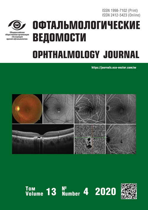
慢性中央浆液性脉络膜病变和卵黄状营养不良成年患者的鉴别诊断方法
- 作者: Matcko N.V.1,2, Gatsu M.V.1,2
-
隶属关系:
- S. Fyodorov Eye Microsurgery Federal State Institution, the Saint Petersburg Branch
- I.I. Mechnikov North-Western State Medical University
- 期: 卷 13, 编号 4 (2020)
- 页面: 35-46
- 栏目: Original study articles
- ##submission.dateSubmitted##: 11.02.2021
- ##submission.dateAccepted##: 16.02.2021
- ##submission.datePublished##: 15.12.2020
- URL: https://journals.eco-vector.com/ov/article/view/60596
- DOI: https://doi.org/10.17816/OV60596
- ID: 60596
如何引用文章
详细
研究目的:优化成年患者发生的慢性中央浆液性脉络膜病变(CSCR)和卵黄状营养不良(VD)的鉴别诊断。
研究任务:在多模式诊断的基础上研究VD和CSCR的特征,通过数学建模来开发诊所不同设施条件下的鉴别诊断方法。
材料和方法:该研究包括61名患有长期神经上皮脱落的患者(90只眼睛)。收集了所有患者的病史,包括家族史,并进行了常规眼科检查:测量最佳矫正视力、裂隙灯检查和眼底照相、视网膜光学相干断层扫描(OCT)和血管造影光学相干断层扫描(OCTA)、短波自发荧光(SW-AF)、视网膜荧光血管造影(FAG)、吲哚菁绿视网膜血管成像(ICGA)。将患者分为2组:30名(30只眼睛)患有卵黄状营养不良的患者和31名(31只眼睛)患有慢性中央浆液性脉络膜病变的患者。采用二元逻辑回归法来评估患病概率。
结果:研究了两组中出现的诊断预测因素,获得了估计检测疾病概率的数学模型。考虑到获得的计算疾病检测概率的公式,包括不同研究组合的标准,制定了鉴别诊断的方法:OCT(曲线下面积0.946);SW-AF(曲线下面积0.955),OCT和SW-AF(曲线下面积0.980);SW-AF、FAG和ICGA(曲线下面积0.989)。
结论:获得的模型可以在不同条件下的诊所对卵黄样营养不良和慢性中央浆液性脉络膜病变进行鉴别诊断。
关键词
全文:
作者简介
Nataliia Matcko
S. Fyodorov Eye Microsurgery Federal State Institution, the Saint Petersburg Branch; I.I. Mechnikov North-Western State Medical University
编辑信件的主要联系方式.
Email: matsko.natalia@mail.ru
ORCID iD: 0000-0001-8909-9999
SPIN 代码: 9790-4066
Ophthalmologist, S. Fyodorov Eye Microsurgery Federal State Institution, Saint Petersburg Branch, PhD Student, I.I. Mechnikov North-Western State Medical University
俄罗斯联邦, St. Petersburg; St. PetersburgMarina Gatsu
S. Fyodorov Eye Microsurgery Federal State Institution, the Saint Petersburg Branch; I.I. Mechnikov North-Western State Medical University
Email: m-gatsu@yandex.ru
Dr. Sci. (Med.), Deputy Director of Clinical Services, S. Fyodorov Eye Microsurgery Federal State Institution, the Saint Petersburg Branch, professor, I.I. Mechnikov North-Western State Medical University
俄罗斯联邦, St. Petersburg; St. Petersburg参考
- Мацко Н.В., Гацу М.В., Григорьева Н.Н. Вителлиформные изменения макулярной области, встречающиеся у взрослых пациентов // Офтальмологические ведомости. – 2019. – T. 12. – № 4. – С. 73–86. [Macko NV, Gacu MV, Grigor’eva NN. Vitelliform changes in the central retina occurring in adults. Opththalmology Journal. 2019;(4):73–86. (In Russ.)] https://doi.org/10.17816/OV18513
- Renner A, Tillack H, Kraus H, et al. Morphology and functional characreristics in adult vitelliform macular dystrophy. Retina. 2004;24(6):929–939. https://doi.org/10.1097/00006982-200412000-00014
- Meunier I, Manes G, Bocquet B, et al. Frequency and Clinical Pattern of Vitelliform Macular Dystrophy Caused by Mutations of Interphotoreceptor Matrix IMPG1 and IMPG2 Genes. Ophthalmology. 2014;121(12):2406–2414. https://doi.org/10.1016/j.ophtha.2014.06.028
- Barbazetto IA, Yannuzzi NA, Klais CM, et al. Pseudo-vitelliform macular detachment and cuticular drusen: exclusion of 6 candidate genes. Ophthalmic Genet. 2007;28(4):192–197. https://doi.org/10.1080/13816810701538596
- Jaouni T, Averbukh E, Burstyn-Cohen T, et al. Association of pattern dystrophy with an HTRA1 singlenucleotide polymorphism. Arch Ophthalmol. 2012;130(8):987–991. https://doi.org/10.1001/archophthalmol.2012.1483
- Wang M, Munch IC, Hasler PW, et al. Central serous chorioretinopathy. Acta Ophthalmol. 2008;86(2):126–145. https://doi.org/10.1111/j.1600-0420.2007.00889.x
- Дога А.В., Качалина Г.Ф., Клепинина О.Б. Центральная серозная хориоретинопатия: современные аспекты диагностики и лечения. – М.: Офтальмология; 2017. – C. 9. [Doga AV, Kachalina GF, Klepinina OB. Central’naja seroznaja horioretinopatija: sovremennye aspekty diagnostiki i lechenija. Moscow: Oftal’mologija; 2017. P. 9. (In Russ.)]
- Lee YS, Kim ES, Kim М, еt al. Atypical vitelliform macular dystrophy misdiagnosed as chronic central serous chorioretinopathy: case reports. BMC Ophthalmology. 2012;12:25. https://doi.org/10.1186/1471-2415-12-25
- Zatreanu L, Freund KB, Leong BCS, еt al. Serous macular detachment in best disease: A masquerade syndrome. Retina. 2020;40(8):1456–1470. https://doi.org/10.1097/IAE.0000000000002659
- Гацу М.В. Фотодинамическая терапия — метод выбора при лечении хронических форм центральной серозной хориоретинопатии // IV Всеросс. семинар-«круглый стол» «Макула-2010»: тезисы докладов. 21–23 мая 2010. – Ростов-на-Дону. – С. 427–429. [Gacu MV. Fotodinamicheskaja terapija – metod vybora pri lechenii hronicheskih form central’noj seroznoj horioretinopatii. IV Vseross. seminar-«kruglyi stol» «Makula-2010»: tezisy dokladov. 21–23 maya 2010. – Rostov-na-Donu, 2010. – P. 427–429. (In Russ.)]
- Балашевич Л.И., Гацу М.В., Искендерова Н.Г. Эффективность диодной субпороговой микроимпульсной лазеркоагуляции при лечении различных форм центральной серозной ретинопатии // IV Всеросс. семинар-«круглый стол» «Макула 2010»: тезисы докладов. 21–23 мая 2010. – Ростов-на-Дону. – С. 416–418. [Jeffektivnost’ diodnoj subporogovoj mikroimpul’snoj lazerkoaguljacii pri lechenii razlichnyh form central’noj seroznoj retinopatii // IV Vseross. seminar-«kruglyj stol» «Makula 2010»: tezisy dokladov. 21–23 maja 2010. – Rostov-na-Donu, 2010. – P. 416–418. (In Russ.)]
- Трухачева H.В. Медицинская статистика в медико-биологических исследованиях пакета Stasistica. – М.: ГЭОТАР-Медиа; 2013. – 379. с. [Truhacheva HV. Medicinskaja statistika v mediko-biologicheskih issledovanijah paketa Stasistica. Moscow: GJeOTAR-Media; 2013. 379. p. (In Russ.)]
- Гонта А. Характеристики изображения: контраст, динамический диапазон, резкость // Алгоритм безопасности. – 2006. – № 5. – С. 56–60. [Gonta A. Harakteristiki izobrazhenija: kontrast, dinamicheskij diapazon, rezkost’. Algoritm bezopasnosti. 2006;5:56–60. (In Russ.)]
- Ergun E. Photodynamic therapy with verteporfin in subfoveal choroidal neovascularization secondary to central serous chorioretinopathy. Arch Ophthalmol. 2004;122(1):37. https://doi.org/10.1001/archopht.122.1.37
- Гацу М.В. Эффективность фотодинамической терапии при хронических формах центральной серозной хориоретинопатии. Причины неудач // VI Всеросс. семинар- «круглый стол» «Макула-2014»: тезисы докладов. 16–18 мая 2014. – Ростов-на-Дону, 2014. – С. 139–155. [Gacu MV. Jeffektivnost’ fotodinamicheskoj terapii pri hronicheskih formah central’noj seroznoj horioretinopatii. Prichiny neudach // VI Vseross. seminar- «kruglyj stol» «Makula-2014»: tezisy dokladov. 16–18 maja 2014. Rostov-na-Donu, 2014. – P. 139–155. (In Russ.)]
补充文件


