


Contemporary aspects of innovative visualization digital medical technologies’ introduction into clinical practice and education

- Authors: Aronov A.M.1, Pastushenko V.L.1, Ivanov D.O.2, Rudin Y.V.3, Drygin A.N.2
-
Affiliations:
- JS LOMO
- St. Petersburg State Pediatric Medical University, Ministry of Healthcare of the Russian Federation
- Institute of Optical-Digital Systems of ITMO University
- Issue: Vol 9, No 4 (2018)
- Pages: 5-11
- Section: Articles
- URL: https://journals.eco-vector.com/pediatr/article/view/10399
- DOI: https://doi.org/10.17816/PED945-11
- ID: 10399
Cite item

Abstract
The paper portrays the elaboration and characters of digital hardware and software complex targeted at automatic forming, registration and processing of biomedical object images for non-invasive diagnostics based upon digital microscopy and endoscopia. Digital hardware and software complex is collecting, preliminary analyzing and compressing video-information for transmission along telecommunication channels. Complexes of this kind may serve as a basis for elaboration and introduction of new prospective medical technologies like visual and digital databases for education programs and atlases for discrimination of pathological cells and states. For a starter an aggregation of clinical and laboratory diagnostics systems has been chosen utilizing images from the outputs of digital microvisual and endoscopical systems for elaboration of optical digital diagnostics complexes. Microvisional digital multispectral analysis system ensures forming and visualization of biological tissues’ and medical slides’ microimages. The videoendoscopic system is intended for endoscopic examination of gastrointestinal tract with forming and visualization of endoscopic images, documenting and archiving of data. This system is networked according to TCP/IP protocol. The microvisional and vi deoendoscopic systems grant distant access to images and control functions for remote users from local networks or through WEB-interface.
Full Text
In recent years, computer and telecommunication technologies have provided a qualitative leap in the development of remote medical technologies. This led to improvement, especially in difficult clinical and diagnostic cases, reliability of diagnosis, and wider coverage of the population with affordable and high-quality diagnostic medical services and ensured reduction in the cost of medical manipulations in pediatrics.
Several leading companies in the world developed and produced automated microimage analyzers, such as Coolscope (Nikon, Tokyo, Japan), BioZero and BioRevo (Keyence, Osaka, Japan), telemedical complexes for ultrasound and x-ray diagnostics, electrocardiography, and computed tomography [7–9]. Portable telemedical terminals have become widespread in the world and enable performance of long-term monitoring of the patient’s cardiovascular system condition, measurement of blood sugar levels, and monitoring other vital indicators of human body homeostasis.According to the World Health Organization, currently hundreds of telemedicine projects have been implemented in the world, which include, besides clinical and informational, educational projects that are also related to tele-education of specialists in the medical field. One problem facingmodern telemedicine is the development of medical informatics methods, standardization of registration and formalization of medical data, and creation of specialized medical institutions capable of providing telemedicine services. To solve these problems, development of information preparation algorithms and their introduction into medical practice are necessary to define standard forms of information exchange at the source data and report generation levels, maintaining medical records in children’s medical institutions and perinatal centers.
In Russia, remote medical technologies have been developing very intensively over recent years. Over the past decade, a coordinating council of the Ministry of Health for telemedicine was organized in our country, the concept of telemedicine technologies development was approved, and the first national standard in the field of medical informatics 1 was developed and adopted, which establishes general provisions for the development of requirements for organizing the creation, maintenance, and use of information systems, such as the electronic health record. New-generation biological digital microscopes, namely, microvizors having advanced telecommunication capabilities, have been developed and mass produced [5].
However, Russian telemedicine hardware lags behind the world level, which is due to the lack of special equipment for clinical and laboratory diagnostics as well as of medical and legal forms for organizing specialized medical institutions capable of providing telemedicine services.
The aforementioned issues, as well as the development of digital and computer technologies in all areas of science and technology, triggered the development of Russian automated telemedicine systems. The first step in this direction was aggregation of clinical and laboratory diagnostics systems that use the medical data as the images obtained from the outputs of digital microvision and video image endoscopy systems, implemented as optical and digital diagnostic complexes for telemedicine (digital diagnostic complexes). Such complexes not only enable expansion of the functionality of existing techniques but also lay the foundation for development and implementation of new promising medical technologies, including creation of visual and digital databases for programs and atlases for recognition of pathological cells and conditions.
The primary aim of the complex developed is creation of an infrastructure basis for development of telemedicine services based on the open information technology of networking of various diagnostic devices. From our viewpoint, they should have the form of advisory and diagnostic centers established on the basis of medical universities, perinatal centers, and leading healthcare facilities of the city and country. These centers will be able to conduct their activities in the form of online consultation and/or based on visual and digital databases.
It is well known that successful treatment of many diseases is determined by an accurate and timely diagnosis. The reliability of diagnostics depends on a large set of various factors, including primarily the qualification of the diagnostician. However, the specialist’s experience and medical intuition alone are not always enough; precise methods and devices for their implementation are required. Long-term professional monitoring of the state of human health and the fullest possible database of various tests are necessary.
Medical records of tests and results of patient examinations in the form of paper-based patient medical records were maintained by doctors for many years, but physical and practical limitations of traditional technologies of storage and organization of a large amount of diversified data are currently completely obvious.
One possible option for the structure of network application of digital diagnostic complexes is presented in Figure 1. The diagram (in the lower part of the figure) shows, as an example, two complexes located in a medical diagnostic facility and connected by a local network to a top-level server of the “electronic hospital” type. Each complex includes three functionally related systems.
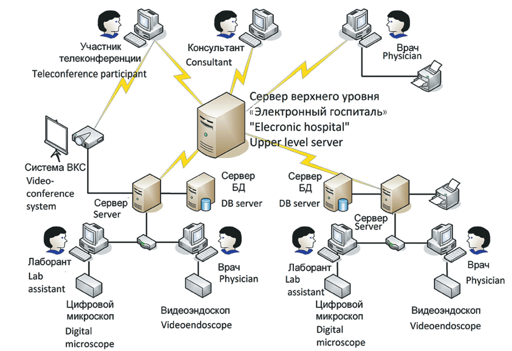
Fig. 1. Network mode diagnostic complex application. LOMO company has elaborated the first multifunctional telemedical diagnostic complex that utilizes the achievements of Russian micro-visional and video-endoscopic techniques in combination with up-to-date computer and telecommunication technologies
Рис. 1. Структура сетевого применения диагностического комплекса. Для этих целей ЛОМО создало первый отечественный многофункциональный диагностический комплекс для телемедицины, использующий достижения отечественной микровизионной и видеоэндоскопической техники в сочетании с современными компьютерными и телекоммуникационными технологиями
The microvisual digital system for multispectral research provides the formation and visualization of microimages of biological tissues and medical preparations.
The video endoscopic system is designed to perform endoscopic examinations of the gastrointestinal tract with the formation and visualization of endoscopic images, documenting and archiving the data.
The network system serves to document and archive the data, compress information for transmission over telecommunication channels, and analyze microimages with the integrated use of data contained in images of various types, based on the use of computer technologies. This system is compatible with modern video-teleconferencing systems, which enables conduction of consultations and case conferences, as well as high-performance exchange of medical data in local, regional, and global telecommunication networks.
DESCRIPTION OF COMPLEX SYSTEMS
The microvision system of a digital diagnostic complex can be characterized as an “all-in-one” system, including a digital microscope, image analyzer, and computer with a network interface in one casing. This system is able to function manually and in automatic mode. It provides the ability to work automatically with samples (scanning across the field, auto focus, change of lighting methods) according to a preset program, save a local copy of the results of microscopic examination, and the possibility to fill in the electronic health record or its integration into existing electronic hospital medical information systems. The microvision system provides remote access to images and control functions for remote users working on a local network or through a web interface.
The main component of the microvision system is an optical and digital microimage analyzer (ODMA). It is a fully automated luminescent microvizor with a built-in integrated control unit based on a personal computer and controllers of actuators with feedback connected to it. ODMA provides microscopic studies in the observing luminescence mode in the visible and near-infrared (IR) spectral ranges, as well as using bright field methods in transmitted, reflected light and under conditions of simultaneous illumination of the objects of observation with transmitted and reflected light of the visible spectrum.
For operation in the automatic mode, ODMA comprises the controlled motorized transfer devices, namely, a two-dimensional microscope stage, focusing mechanism, translucent light ray filter unit, dia-illuminator diaphragm, objective turret, unit of beam-splitting modules of reflected light, node for switching on the IR channel of the reflected light, node for switching on the light-emitting diode (LED) illuminator of the reflected light, valve, and device for transfer of the mercury lamp collector. In the remote control mode, the main functions of ODMA support the transmissioncontrol protocol/Internet protocol (TCP/IP) network protocol.
Due to the ODMA software, microimages are recorded, including the construction of panoramic X–Y images with automatic linking of field boundaries and Z-scanning with recording of images in “deep focus” mode, as well as their preliminary processing, compression, and transfer for archiving to the network system of the complex. To obtain the best quality microimages, the ODMA software implements an automatic assessment of the contrast and sharpness of digital images and also supports the automatic focusing mode, which algorithms and basic parameters were studied previously [2–4].
The video endoscopic system of a digital diagnostic complex serves as the workplace of an endoscopist and includes a video endoscope with a tool kit, lighting unit, control unit, video monitor, and personal computer with software installed on the instrument endoscopic rack.
The video endoscopic system was created using a number of new technical solutions aimed primarily at improvement of the image quality, as well as of consumer properties and performance characteristics. The image receptor in a video endoscope is used as a 1/6 inch color CCD matrix with a pixel size of 3.275 × 3.150 μm, for which a new lens 2 with a 140° angular field of view was developed. This lens provides high-quality color images of the object across the entire field without refocusing at working distances of 3 to 100 mm, the distribution of illumination across the image field (uneven illumination does not exceed 25%).
Figure 2 shows the optical scheme of the lens, and Table 1 represents the calculated values of the image contrast transfer coefficient for different working distances and spatial frequencies.

Fig. 2. Video-endoscope objective optical scheme
Рис. 2. Оптическая схема объектива видеоэндоскопа
Table 1. Contrast transfer design coefficient value
Таблица 1. Расчетные значения коэффициента передачи контраста изображения
Operative distance, mm / Рабочее расстояние, мм | Contrast transfer design coefficient value for field center at spatial frequency / Расчетное значение коэффициента передачи контраста для центра поля на пространственной частоте | ||
40 mm–1 | 50 mm–1 | 110 mm–1 | |
3 | 0.50 | 0.38 | – |
4.5 | – | 0.61 | – |
12 | – | 0.59 | 0.27 |
100 | – | 0.65 | 0.33 |
In addition, the controlling mechanism of the bendable part of the video endoscope and braking devices is hermetically sealed, the ergonomic characteristics of the proximal part and control handles are improved, and the shape of all structural elements provides comfortable working conditions for doctors with various anthropometric data. LED illumination is provided in the lighting channel of the video endoscopic system. The required illumination is achieved using a super-bright white LED as a light source, wide-angle lighting lenses, and fiber optic bundles with increased transmission (Fig. 3).

Fig. 3. Optical digital micro-image analyzer optical scheme: 1 – changeable micro-objectives block; 2 – spectro-diviser block; 3 – photo-receiver block; 4 – transmitted light LED; 5 – Hg-lamp lighthead, 6 – reflected light LED; 7 – laser lighthead
Рис. 3. Оптическая схема оптико-цифрового анализатора микроизображений: 1 — блок сменных микрообъективов; 2 — блок спектроделителей; 3 — блок фотоприемников; 4 — светодиодный осветитель проходящего света; 5 — осветительный модуль ртутной лампы; 6 — светодиодный осветитель отраженного света; 7 — лазерный осветительный модуль
The control unit of the video endoscopic system was upgraded to meet the requirements of improved image quality and compatibility with the network system of the complex. For this purpose, using the menu commands in the control unit, the functions of controlling color grade, clarity, and brightness of the image are implemented. To optimize the observation mode in the process of endoscopic examination, it is possible to change the size of the angular field of vision of the lens using an electronic mask and display a still image simultaneously with the video image (picture-in-a-picture mode). Communication with the network system is supported by TCP/IP.
Thus, the first Russian digital video endoscope for telemedicine was created, which provides the opportunity to monitor endoscopic procedures remotely during their execution.
In addition to the diagnostic functions performed by microvision and video endoscopic systems, which are important from the medical use viewpoint, digital diagnostic complexes include a network system for solving telemedical problems. This system is a software and hardware complex deployed on the basis of the HP ProLiant ML150 G6 server and supporting software for managing the database of diagnostic studies coming to the server from the diagnostic systems of the complex using the Digital Imaging and Communications in Medicine protocol, which is the industry standard for creating, storing, transmitting, and visualizing medical images and documents of the patients examined. Reliability of diagnostics in observable images is ensured by inclusion of a number of original computer programs for medical image processing into the program complex of the network system [1, 6]. The network system of the digital diagnostic complex is an open information system capable of supporting software products from other manufacturers. With its application, remote users, in and outside of the local network of the complex, subject to authorization, can access the control functions of the microvision system and monitor streaming video coming from the video endoscopic system, as well as personal electronic medical records of patients stored in a database of diagnostic studies. The ability to connect to the Internet and the telecommunication functions of the network system itself define strict requirements for the protection of personal data and information security of the complex as a whole.
CONCLUSIONS
As a result of the work in a joint project of the LOMO company and St. Petersburg National Research University of Information Technologies, Mechanics and Optics, the first Russian high-tech optical and digital diagnostics complex for telemedicine was developed. The complex was designed to conduct clinical and laboratory research and to solve a relevant objective of improving quality of medical care for broad layers of the Russian population, including those living in areas remote from modern diagnostic centers. The open network architecture provides for expansion of the scope of application of the complex in medical practice due to including new diagnostic tools in its composition (Fig. 4).

Fig. 4. Micro-image optical digital analyzer
Рис. 4. Внешний вид оптико-цифрового анализатора микроизображений
This work was supported by the Ministry of Education and Science of the Russian Federation.
About the authors
Alexander M. Aronov
JS LOMO
Author for correspondence.
Email: lomo@lomo.sp.ru
Dr Sci, Professor, Director
Russian Federation, Saint PetersburgVladimir L. Pastushenko
JS LOMO
Email: lomo@lomo.sp.ru
Dr Sci, Professor, Director General’s Advisor
Russian Federation, Saint PetersburgDmitry O. Ivanov
St. Petersburg State Pediatric Medical University, Ministry of Healthcare of the Russian Federation
Email: doivanov@yandex.ru
MD, PhD, Dr Med Sci, Professor, Rector
Russian Federation, Saint PetersburgYaroslav V. Rudin
Institute of Optical-Digital Systems of ITMO University
Email: yaroslav-r@mail.ru
PHD, Associate Professor, Director
Russian Federation, Saint PetersburgAlexey N. Drygin
St. Petersburg State Pediatric Medical University, Ministry of Healthcare of the Russian Federation
Email: 9112286592@mail.ru
MD, PhD, Dr Mеd Sci, Professor, Head, Research Center
Russian Federation, Saint PetersburgReferences
- Аверкин А.Н., Потапов А.С. Применение метода восстановления глубины из фокусировки для микроскопических изображений // Оптический журнал. - 2011. - Т. 78. - № 11. - С. 52-59. [Averkin AN, Potapov AS. Using the method of depth reconstruction from focusing for microscope images. Opticheskiy zhurnal. 2011;78(11):52-59. (In Russ.)]
- Беззубик В.В., Белашенков Н.Р., Устинов С.Н. Оптимизация алгоритмов автофокусировки цифрового микроскопа // Оптический журнал. - 2009. - Т. 76. - № 10. - С. 16-22. [Bezzubik VV, Belashenkov NR, Ustinov SN. Optimization of algorithms for autofocusing a digital microscope. Opticheskiy zhurnal. 2009;76(10):16-22. (In Russ.)]
- Белашенков Н.Р., Беззубик В.В., Никифоров В.О. Анализ влияния дефокусировки и шума на качество цифрового изображения // Научно-технический вестник Санкт-Петербургского государственного университета информационных технологий, механики и оптики. - 2011. - № 6. - С. 59-64. [Belashenkov NR, Bezzubik VV, Nikiforov VO. Analysis of blur and noise influence on digital image quality. Nauchno-tekhnicheskiy vestnik Sankt-Peterburgskogo gosudarstvennogo universiteta informatsionnykh tekhnologiy, mekhaniki i optiki. 2011;(6): 59-64. (In Russ.)]
- Белашенков Н.Р., Беззубик В.В., Никифоров В.О. Метод количественной оценки контраста цифрового изображения // Научно-технический вестник Санкт-Петербургского государственного университета информационных технологий, механики и оптики. - 2010. - № 6. - С. 86-88. [Belashenkov NR, Bezzubik VV, Nikiforov VO. Quantitative estimation method of digital image contrast. Nauchno-tekhnicheskiy vestnik Sankt-Peterburgskogo gosudarstvennogo universiteta informatsionnykh tekhnologiy, mekhaniki i optiki. 2010;(6):86-88. (In Russ.)]
- Белашенков Н.Р., Калинина Т.Ф., Лопатин А.И., и др. Микровизоры - новое поколение цифровых микроскопов // Оптический журнал. - 2009. - Т. 76. - № 10. - С. 52-57. [Belashenkov NR, Kalinina TF, Lopatin AI, et al. Microviewers - the next generation of digital microscopes. Opticheskiy zhurnal. 2009;76(10):52-57. (In Russ.)]
- Дырнаев А.В. Метод подсчета эритроцитов на изображениях мазков крови // Научно-технический вестник Санкт-Петербургского государственного университета информационных технологий, механики и оптики. - 2011. - № 2. - С. 17-22. [Dyrnaev AV. Red cells count method on blood smears images. Nauchno-tekhnicheskiy vestnik Sankt-Peterburgskogo gosudarstvennogo universiteta informatsionnykh tekhnologiy, mekhaniki i optiki. 2011;(2):17-22. (In Russ.)]
- Falconer J, Giles W, Villanueva H. Realtime ultrasound diagnosis over a wide-area network (WAN) using off-the-shelf components. J Telemed Telecare. 1997;3 Suppl 1:28-30. doi: 10.1258/1357633971930265.
- Thrall JH, Boland G. Telemedicine in practice. Semin Nucl Med. 1998;28(2):145-157. doi: 10.1016/s0001-2998(98)80004-4.
- Tsagaris MJ, Papavassiliou MV, Chatzipantazi PD, et al. The contribution of telemedicine to cardiology. J Telemed Telecare. 1997;3 Suppl 1:63-64. doi: 10.1258/1357633971930418.
Supplementary files

