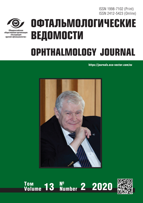
卷 13, 编号 2 (2020)
- 年: 2020
- ##issue.datePublished##: 24.08.2020
- 文章: 12
- URL: https://journals.eco-vector.com/ov/issue/view/1434
- DOI: https://doi.org/10.17816/OV20202
Obituary
In memory of Yuri Sergeevich Astakhov
摘要
He passed a long and bright path, A worthy person. He did not betray, he did not lose heart And disappeared forever ... How difficult it is for you and me to accept that he will never send a smile in return, he will not ask: "How are you?" That there was a AMAZING MAN next to us in life!
 6-8
6-8


Original study articles
鼻内窥镜术对提高慢性泪囊炎患者手术疗效的条件
摘要
前言:慢性泪囊炎患者的外科治疗是一个不简单的跨学科难题。通往泪囊的外路和内路是众所周知的。治疗效果在很大程度上取决于全面的诊断和治疗方法的选择。
目的:鼻内入路手术治疗慢性泪囊炎的疗效分析。
材料和方法:2015-2019年期间对225例慢性泪囊炎患者的研究。将患者分为2组:第一组(110例患者)进行了鼻内镜下泪囊鼻腔吻合术(EEDCR),第二组(115例患者)进行了鼻内镜下泪囊鼻腔吻合术(EEDCR)并辅以鼻内干预。所有患者均接受了鼻腔内窥镜检查和脸部中央区域的MSCT检查。
结果:所得数据结果显示,与鼻内镜下泪囊鼻腔吻合术(EEDCR)相比,对已确定鼻内定位病变行联合手术
(EEDCR+)的患者组,其疗效更高。
结论:根据该研究,患有慢性泪囊炎的患者必须进行全面的眼科及鼻内诊断,包括脸部中央区域的MSCT泪囊造影检查。当病理过程导致难以进行常规EEDCR时,有必要扩大手术范围,使慢性泪囊炎患者的疗效提高至91.3%。
 15-21
15-21


Optimization of energy parameters during surgery of high-density cataract
摘要
The goal is to develop a combined technique for preliminary YAG laser fragmentation and Femto laser exposure on a CATALYS device and to evaluate its role in reducing the time and energy parameters of a surgery of high-density cataract.
Material and methods. The study included 118 patients (118 eyes) with age-related cataracts of the 3rd and 4th degrees of lens nucleus density. In the main group, before phacoemulsification (PE) with a Femto laser support and IOL implantation, preliminary YAG laser phacofragmentation of the lens nucleus was performed. In the first control group, PE was performed with Femto laser support and IOL implantation. In the second control group – PE with IOL implantation.
Results. A 35% decrease in the energy of the Femto laser action at a 3rd degree of lens nucleus density was achieved, and 40% – at the 4th degree, in comparison with the PE with the Femto laser support without preliminary YAG laser phacofragmentation, a 38% decrease in the cumulative ultrasound energy at 3rd degree of the lens nucleus density, and 42% at 4th degree compared with isolated ultrasonic cataract phacoemulsification.
Conclusion. The proposed modification of the technique of combined YAG laser and Femto laser exposure allows achieving during cataract surgery a complete fragmentation of the lens nucleus of a high degree of density, helps minimizing the risk of complications and reaching quick postoperative rehabilitation of patients.
 23-30
23-30


黄斑水肿的EN FACE光学相干断层成像、视网膜厚度图及荧光血管造影图像的代表性
摘要
目的:研究黄斑水肿的en face光学相干断层成像、视网膜厚度图及荧光血管造影图像的代表性。
材料和方法:在这项回顾性横断面研究中包括8例(15只眼)糖尿病性黄斑水肿患者(其中2名女性和6名男性,平均年龄为66.5 ± 8.1岁)。所有患者均接受了OCT(厚度图和en face图像)和荧光血管造影检测。en face OCT的视网膜层图像是从内部丛状层向外产生。2名研究员独立评估了水肿面积。
结果:根据荧光血管造影、en face OCT以及视网膜厚度图像的评估结果,视网膜水肿区的指标分别为12.7 ± 8.1, 14.5 ± 8.4, 10.4 ± 6.9 mm2 (ANOVA × 3, p = 0.34),在统计学上无显著差异。三种方法及单独平均测量的组内相关系数分别为 0.87 和 0.95。
结论:对黄斑水肿区的评估以及黄斑区“格子样”激光治疗的规划,en face OCT是取代荧光血管造影检测非常合适的非侵入性检测方法。
 9-14
9-14


急性角膜水肿:诊断和治疗
摘要
急性角膜水肿是一种以后弹力膜破裂导致的角膜基质水肿为特征的疾病。
研究目的:分析急性角膜水肿患者的诊断和治疗结果。
材料和方法:该研究组中包括42例(47只眼)急性角膜水肿患者。5例患者两只眼同时或先后发生急性角膜水肿。平均年龄为28.7 ± 10.1岁(19岁到54岁),男31例,女11例,观察期为5年。在没有药物治疗作用或术后
并发症的情况下:向眼前房注入10%气体(C3F8, SF6),羊膜移植,板层角膜移植,深层前角膜移植,穿透性角
膜移植。
结果:急性角膜水肿发作前的平均病程为12.6 ± 4.6年。11.9%的患者先前未诊断出角膜扩张。局部水肿的角膜厚度为692 ± 98 μm,全水肿的角膜厚度为1200 ± 220 μm。与局部和部分水肿相比,次全和全角膜水肿其损伤程度、后弹力层脱离的高度以及破裂边缘分叉更明显(χ2, p < 0.001)。将10%的气体-空气混合物(C3F8, SF6)注入部分和次全角膜水肿患者的眼前房,可以明显加快水肿缓解。
结论:这类病例的分析结果表明,根据急性角膜水肿的严重程度采用不同的治疗方案。
 31-42
31-42


后期圆锥角膜后弹力层基质内移植
摘要
目的:后期圆锥角膜患者后弹力膜(DM)基质内移植术后观察结果报告。
方法:对三名进行性圆锥角膜患者(3只眼)进行了检查。患者之前没有接受过紫外线交联或任何类型的眼科手术。所有患者均在局部麻醉下接受了DM移植入基质内。手术治疗经过12个月后进行了临床结果评估。
结果:根据我们的资料,这是有关基质内DM移植的第一份报告。文献中也没有关于进行性圆锥角膜患者使用DM移植替代Bowman层移植的数据。手术12个月后,DM移植物在角膜基质中没有褶皱,几乎看不见,而且角膜透明。未发现裸眼视力(UCVA)、隐形眼镜(CL)的最佳矫正视力(BCVA)、内皮细胞密度(ECD)、最小角膜厚度(minTP)和最大角膜曲率指数(Kmax)的显著变化。术中或术后未观察到并发症。
结论:DM的基质内移植可以替代后期圆锥角膜患者Bowman层的移植,这可以避免或延缓进行PSC(穿透性角膜移植术)或DALK(深层前角膜角膜移植术)角膜移植术。
 43-48
43-48


Reviews
Modern principles of the diabetic macular edema management
摘要
Diabetes mellitus and diabetic retinal lesions are a global challenge for healthcare systems and one of the leading causes of severe vision loss among the working age population. Retinal laser coagulation remained the standard of therapy and the only possible treatment for diabetic macular edema (DME) in the 80-90s of the last century. The introduction of anti-VEGF therapy and glucocorticoids into the wide practice has significantly expanded the range of possibilities of DME treatment, allowing not only to stabilize patients’ vision, but also to improve it. The analyses of the large randomized clinical trials results are made and presented in this article, that highlight the basic principles of the contemporary DME treatment. This information is intended to help the ophthalmologist to develop the most optimal approach to treatment, considering the individual characteristics of each patient and the “evidence-based” efficacy and safety data of different methods.
 51-65
51-65


Selective laser trabeculoplasty: mechanisms of action and predictors of efficacy
摘要
This review considered selective laser trabeculoplasty in comparison with other methods for therapeutic and surgical treatment of primary open-angle glaucoma and ocular hypertension, including argon laser trabeculoplasty. In this paper, we reviewed current knowledge on the mechanisms of action of selective laser trabeculoplasty, predictors of intraocular pressure-lowering effect, repeatability, and safety of this procedure.
 67-76
67-76


Lamellar macular holes: the evolution of ideas about pathogenesis and the clinical picture. The development of diagnostic approaches. Modern classification
摘要
This review presents the development of modern ideas about the pathogenesis of lamellar macular holes, anatomical changes, as well as the formation of a modern classification based on the expansion of diagnostic capabilities, and differences from other similar conditions of the vitreomacular interface. An evolutionary breakthrough in the investigation of this disease was associated with the widespread introduction of optical coherence tomography, while the development of its resolution allowed us to detect some features of epiretinal proliferation that occurs in lamellar macular holes, which are also addressed in the review.
 77-88
77-88


Case reports
Bilateral lacrimal gland enlargement as a clinical manifestation of sarcoidosis
摘要
Sarcoidosis is an inflammatory disorder of unknown etiology. The main characteristics of sarcoidosis are non-caseating granulomas which can be formed in affected organs. Eye and its adnexal tissues can also be involved. In the early stages of sarcoidosis, the clinical picture may be mild and not always specific. Although the early signs of sarcoidosis are lung associated, we present a clinical case with diffuse enlargement of the lacrimal glands as a first sign of systemic sarcoidosis. A 21-years-old woman presented to the ophthalmological center with a 2-weeks history of having swollen, painful upper eyelids. Primary eye examination revealed palpable tender masses in the locations of the lacrimal glands. Characteristic for sarcoidosis X-ray findings and blood tests demanded lacrimal gland and lung biopsy for diagnosis verification. In spite sarcoidosis of lacrimal glands is a rare condition, it has to be taken into consideration in the differential diagnosis of lacrimal gland masses.
 89-94
89-94


Complex clinical case of optic neuropathy differential diagnosis
摘要
The outcome of any damage or disease of the optic nerve is the atrophy of the optic nerve, which causes the irreversible loss of vision. The reasons of damage of the optic nerve are polymorphous, and early diagnosis allows to improve the efficacy of treatment and prevent irreversible consequences. The article presents a difficult clinical case of optic neuropathy differential diagnosis.
 95-99
95-99


Recurrent pterygium – features of surgical treatment
摘要
Pterygium is a fibrovascular degenerative condition of the subconjunctival tissue that proliferates and grows into the cornea in the form of a vascularized fold, destroying the surface layers of the stroma and Bowman’s membrane. This disease is common throughout the world. The etiology is not clear, but it is known that the appearance of pterygium is associated with exposure of the eye to ultraviolet rays. The treatment of the de novo occurring pterygium is a relatively simple task and involves surgical removal. But a simple excision is currently unacceptable due to the high recurrence rate. In order to minimize the risk of relapse, numerous adjuvant treatment methods are used, which include anti-metabolites such as Mitomycin C and 5-fluorouracil, amniotic membrane, various types of conjunctival and or limbal grafts; medications such as anti-vascular endothelial growth factor are sometimes used. In the clinical case presented in the article, we successfully used the technique of anterior lamellar keratoplasty and autoconjunctival transplantation in combination with intra-operative use of anti-metabolite Mitomycin C in a young patient with recurrent stage IV pterygium (degree of activity 3) twice unsuccessfully operated on. As a result of this surgical technique, the transparency of the cornea was restored and high visual acuity was obtained.
 101-107


