Vol 12, No 1 (2019)
- Year: 2019
- Published: 06.06.2019
- Articles: 12
- URL: https://journals.eco-vector.com/ov/issue/view/695
- DOI: https://doi.org/10.17816/OV20191
Original study articles
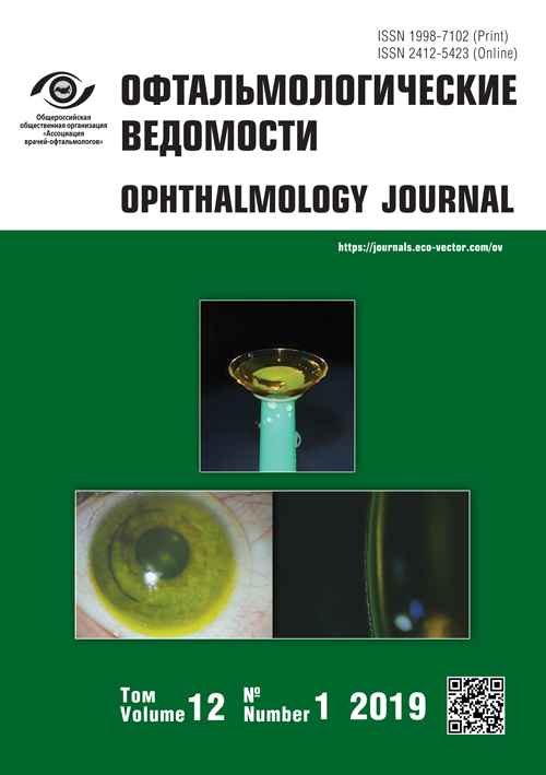
Miniscleral lenses in the treatment of patients with dry eye syndrome (first own experience)
Abstract
Background. Scleral lenses, due to their benefits, hold a specific position among all types of contact lenses. Some years ago, they began to be used successfully not only for the correction of complex types of refractive errors, when other types of correction failed to achieve satisfactory visual function and visual rehabilitation of patients, but also as a therapeutic system in the management of ocular surface disease.
Purpose. To evaluate the efficacy of rigid gas permeable miniscleral contact lenses as a therapeutic system in the management of patients with dry eye syndrome by filling the space under the lens with a non-preserved sodium hyaluronate solution.
Materials and methods. In the study, 7 patients (11 eyes) with keratectasias after corneal surgery and concomitant dry eye syndrome were included. In the treatment and rehabilitation of these patients, miniscleral contact lenses were used during daytime with additional filling of the space under the lens with a non-preserved sodium hyaluronate solution.
Results. As a criterion of the effectiveness of miniscleral contact lens use for therapeutic purposes, along with a significant increase in visual function in patients with complex corneal pathology, the elimination of discomfort due to restoration of the corneal epithelium integrity and improvement of their quality of life is considered.
 5-12
5-12


Artificial intelligence and machine learning for optical coherence tomography-based diagnosis in central serous chorioretinopathy
Abstract
The aim of the present study was to examine the potential of machine learning for identification of isolated neurosensory retina detachment and retinal pigment epithelium (RPE) alterations as diagnostic criteria of central serous chorioretinopathy (CSC).
Material and methods. Patients with acute CSC in whom a standard ophthalmic examination and optical coherence tomography (OCT) using RTVue-XR Avanti (Angio Retina HD scan protocol, 6 × 6 mm) was performed were included in the study. 10-μm en face slab above the RPE layer was used to create ground truth masks. Learning aims were defined as identification of 3 classes of structural abnormalities on OCT cross-sectional scans: class 1 – subretinal fluid, class 2 – RPE abnormalities, and class 3 – leakage points. Data for each of the 3 classes included: 4800/1400 training/test images for class 1, 2000/802 training/test images for class 2, and 1504/408 training/test images for class 3. Unet-similar architecture was used for segmentation of abnormalities on OCT cross-sectional scans.
Results. Analysis of test sets revealed sensitivity, specificity, precision, and F1-score for detection of subretinal fluid of 0.61, 0.99, 0.99, and 0.76, respectively. For detection of RPE abnormalities sensitivity, specificity, precision, and F1-score were 0.14, 0.95, 0.94 and 0.24, respectively. For detection of leakage point sensitivity, specificity, precision, and F1-score were 0.06, 1.0, 1.0, and 0.12, respectively.
Conclusions. Thus, machine learning demonstrated high potential in the OCT-based identification of structural abnormalities associated with acute CSC (neurosensory retina detachment and RPE alterations). Topical identification of the leakage point appears to be possible using large learning sets.
 13-20
13-20


Anatomic features of anterior chamber angle in children with glaucoma depending on the degree of the cicatricial retinopathy of prematurity
Abstract
Introduction. The retinopathy of prematurity (ROP) is a leading condition in the nosological structure of ophthalmic pathology in preterm children. A number of researchers note the increase in frequency of glaucoma development in such patients, considerably worsening the prognosis of the disease. At the same time, features of ocular hydrostatics and hydrodynamics taking into account the immaturity of the eye are studied insufficiently.
The purpose of the study was to estimate the anterior chamber angle anatomy in preterm children with glaucoma depending on the cicatricial ROP severity.
Materials and methods. The study group included 45 preterm children (87 eyes) aged from 6 months to 18 years with glaucoma on the background of cicatricial ROP. The control group consisted of 27 full-term children (54 eyes) with congenital glaucoma. As an addition to traditional ophthalmologic examination, iridocorneal goniography using a pediatric retinal camera was performed.
Results. In children of the study group, anomalies of anterior chamber angle anatomic structure were diagnosed in the absolute majority of cases – 97.7% of cases (85 eyes) and depended on the cicatricial ROP severity: at degrees 1-3 – they were similar to those in congenital glaucoma in full-term children; at degrees 4-5 – glaucoma was of secondary nature and developed as a result of a combination of dysgenesis signs in the anterior chamber angle.
Conclusion. Cicatricial ROP of any degree is a high risk factor for the development of secondary glaucoma.
 21-26
21-26


Intravitreal injections in clinical practice: results of a survey of eye surgeons in the Ural federal district
Abstract
Introduction. Intravitreal i njections ( IVI) are widespread in modern ophthalmology as a method of drug delivery in various posterior segment pathologies. Despite the accumulated experience, some aspects of the procedure remain debatable. In addition, in the literature there are no data on the practical aspects of IVI performance in real clinical conditions in Russia. This article presents the results of an anonymous survey of 74 ophthalmologists of the Ural Federal District of the Russian Federation, conducted in 2018 using an original questionnaire, which included 20 questions. It was revealed that respondents generally share the traditional for Russia approach to IVI as to major ophthalmic surgery. The survey revealed a lack of common approaches and incomplete adherence to modern guidelines in certain aspects of the procedure, and therefore correcting measures were proposed.
 27-36
27-36


Reviews
Modern notions about the tactics of treatment of patients with non-full thickness macular holes: to observe or to operate?
Abstract
In this review, the opinions of different authors on the problem of non-full thickness macular holes are discussed in detail. Currently, there are three different approaches to the management of this condition. Dynamic observation allows assessing the degree of their progression, to determine some or other anatomical indicators which influence the functional state of the retina and visual function. Pharmacological vitreolysis in some cases allows eliminating vertical and tangential traction in a least invasive mannor. To resolve this problem in a radical way is possible by surgical treatment – posterior vitrectomy, but this is also related to certain surgical risks, and does not always lead to an increase in visual acuity. As a rule, it is recommended to patients with a significant decrease in visual acuity. Currently, indications for surgical treatment of patients with high visual function are ambiguous.
 37-44
37-44


Algorithm of objective examination of a patient with blepharoptosis
Abstract
Meticulous objective examination of a patient with blepharoptosis allows determining the tactics of surgical treatment. It depends on many factors, but main ones are blepharoptosis etiology, upper eyelid’s levator function, and ptosis degree. The estimation algorithm of objective examination of a patient with blepharoptosis is presented in this article.
 45-51
45-51


Ophthalmopharmacology
Features of the epithelial layer formation on corneal graft
Abstract
Relevance. Significant impact on graft survival has a delay in the formation of epithelial cover.
Purpose – cytological characteristics and analysis of the effect of medications on the epithelial layer formation on the graft.
Materials and methods. An analysis of the post-operative state of 20 patients operated for bullous keratopathy and corneal ulcer. Smears from the surface of the graft were stained by Leucodif 200 (LDF 200) and examined by light microscopy.
Results. The epithelial layer formation in all cases ended no later than 8-10 days after keratoplasty. According to the cytological analysis, a complex of postoperative treatment which included Vixipin from the 10th day of the postoperative period. Visual acuity improvement occurred in 11 patients (75% of cases) operated due for bullous keratopathy, and in four patients operated for corneal ulcer.
Conclusions. The epithelial layer formation on the surface of corneal transplant occurs due to the migration of mature epithelial cells from the wound edges of the own cornea and mitotic activity of limbal cells of the recipient. Long-lasting epithelial defect and inflammation preceding keratoplasty result in slower epithelialization of the graft in the postoperative period. Use of corticosteroids, sodium hyaluronate and antioxidants in the postoperative period contribute to the formation of a full epithelial layer.
 53-58
53-58


Lectures
Differential diagnosis of optic neuropathy in patients with CNS diseases and normal tension glaucoma
 73-76
73-76


In ophthalmology practitioners
Acanthamoeba keratitis. Review of literature. Case reports
Abstract
Acanthamoeba keratitis (АК) is a parasitic infectious condition caused by corneal invasion by free-living amoebae. In 86% of cases, AK affects contact lens wearers. Delayed diagnosis and inadequate treatment of this disease leads to development of a severe form of keratouveitis and corneal perforation. Consequently, this group of diseases is one of the causes of visual disability in working-age population.
 59-71
59-71


Case reports
Optical coherence tomography in optic disc pit diagnosis
Abstract
The optic disc pit is a congenital anomaly, presenting as a round or oval depression in the optic disc, 0.1–0.7 disc diameters in size, located mainly along its lower-temporal margin. Approximately in 45-75% of the eyes with congenital optic disc pit a serous detachment of neuroepithelium (NED) in the macular area develops, which could become a cause for an erroneous diagnosis of an intraocular tumor. The article presents a clinical case of the optic disc pit, which required a differential diagnosis with an intraocular tumor. It is shown that OCT signs of the optic disc pit are: the connection of optic disc structures with the subretinal space, as well as the presence of signs of invagination of the retinal nerve fiber layer in the optic disc structures.
 77-82
77-82


Experience in navigation system use in surgical treatment of the traumatic dacryocystitis recurrence (clinical case)
Abstract
The article presents a clinical case of surgical of a recurrent chronic purulent dacryocystitisof traumatic etiology. Under endoscopic control, the patient underwent a revisional endonasal laser dacryocystorhinostomy with bicanalicular silicone stent. All surgical steps were controlled on the navigation system display. Navigation equipment was used to accurately visualize the structures of the nasal cavity and lacrimal pathways, because the anatomy of this area was changed as a result of the midface trauma and preliminary unsuccessful surgical procedures. During surgery, the functional and anatomical patency of the lacrimal pathways was achieved. Thus, the navigation system is a useful supplementary equipment that allows identifying the lacrimal sac in complex surgical situations, to monitor the state of lacrimal passages and surrounding nasal structures, which ultimately increases the effectiveness and safety of the procedure.
 83-88
83-88


Obituary
 89-93
89-93


