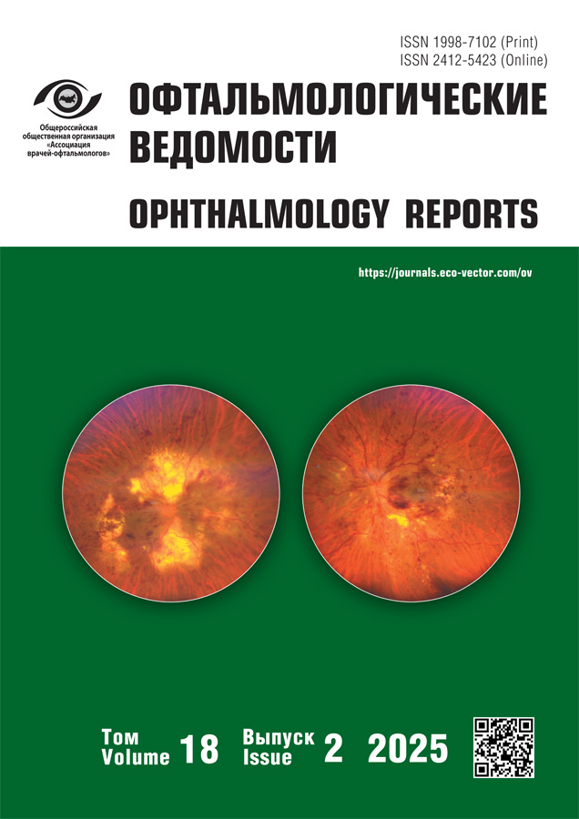
Ophthalmology Reports
Medical peer-reviewed quarterly journal published since 2008.
Publisher
- Eco-Vector
WEB: https://eco-vector.com/
Chief editor
- professor Dmitriy V. Davydov, MD, Dr. Sci. (Medicine)
ORCID: 0000-0001-5506-6021
About
Main publications of the journal are focused on key issues of modern ophthalmology: etiology and pathogenesis, epidemiology, clinical picture features, up-to-date methods of diagnosis, prevention, and treatment of eye diseases and of those of its adnexa.
The journal publishes original articles, scientific reviews, lectures, clinical case descriptions (presented by Russian and foreign authors), and informs about past congresses and conferences in Russia.
The journal is oriented toward practicing ophthalmologists, including ophthalmic surgeons, scientific and teaching staff of medical higher educational institutions, physicians in ophthalmology training, as well as for specialists of allied health specialties.
The journal’s mission:
- To integrate research results of Russian scientists and the rich clinical experience of practicing doctors in diagnosis, prevention, and treatment of eye diseases into the international scientific space; to be an international scientific platform for discussions and sharing experiences;
- To provide for ophthalmologists of the Russian Federation actual and high quality research and practice insights into most up-to-date treatment and prevention methods of eye diseases and of those of its adnexa.
Publications
- in English, Russian, Chinese
- in hybrid access (subscription and Open Access with СС BY-NC-ND license)
- with no obligatory APC for all authors
Indexation
- elibrary
- BASE
- Crossref
- Dimensions
- Fatcat
- Google Scholar;
- OpenAlexScilit
- RSCI
- Scholia
- Scopus
- Ulrich's Periodicals directory
- Wikidata
Current Issue
Vol 18, No 2 (2025)
- Year: 2025
- Published: 18.07.2025
- Articles: 11
- URL: https://journals.eco-vector.com/ov/issue/view/10239
- DOI: https://doi.org/10.17816/OV20252
Original study articles
Prognostic value of BAP1 protein expression in uveal melanoma
Abstract
BACKGROUND: Uveal melanoma is the most common malignant ocular tumor in adults. It carries a high risk of metastatic spread and death. Typical clinical and morphological signs fail to provide accurate disease prognosis. Thus, investigations of molecular markers such as BAP1 expression are warranted to improve survival prediction and optimize treatment strategies.
AIM: The work aimed to determine the prognostic value of the histological type of uveal melanoma and BAP1 expression for survival of patients.
METHODS: We performed a retrospective analysis of the data of 68 patients with uveal melanoma who received curative treatment. A standard procedure was used for the morphological examination of enucleated eyes. BAP1 protein expression was evaluated using immunohistochemistry. Survival was analyzed using Kaplan–Meyer methods and a Cox proportional hazard model.
RESULTS: Median survival in patients with homo- or heterogeneous (focal, mosaic) loss of BAP1 expression was 48 months, whereas patients with homogeneous BAP1 expression of variable degree (mild to severe) did not achieve the median by the end of follow-up. The log-rank test showed statistically significant differences between these groups (χ2=4.344; p=0.037). Mortality risk for patients with homo- or heterogeneous loss of BAP1 expression was 2.6 times higher (HR=2.602, 95% confidence interval: 0.573–0.96). However, mortality risk for patients with epithelioid cell and mixed tumor types was only 1.27 times higher than for patients with spindle cell cancer (HR=1.265, 95% confidence interval: 1.062–2.846).
CONCLUSION: The study highlights the importance of using molecular genetic methods, including immunohistochemistry of BAP1, to predict disease outcomes more accurately.
 7-16
7-16


Specific clinical manifestations of proliferative diabetic retinopathy in young patients and assessment of technical challenges of endovitreal surgery and its outcomes
Abstract
BACKGROUND: The number of young patients with type 1 diabetes mellitus is steadily increasing in all countries of the world. Technical features of performing vitrectomy for proliferative diabetic retinopathy in patients with type 1 diabetes mellitus have not been sufficiently studied. The need for their study is very urgent, since the number of such patients is constantly increasing, the information obtained will help to avoid intra- and postoperative complications that may arise during vitrectomy.
AIM: The work aimed to study the morphological and functional features of proliferative diabetic retinopathy in young patients with type 1 diabetes mellitus and to assess the technical challenges of endovitreal surgery and its outcomes.
METHODS: The study included unselected young patients with proliferative diabetic retinopathy and type 1 diabetes mellitus who were indicated for vitreoretinal surgery. A total of 32 patients (55 eyes) aged 18 to 46 years were selected; best corrected visual acuity with light projection was up to 0.3. A three-port pars plana endovitreal procedure was performed in all patients.
RESULTS: A total of 48 eyes had dense fused posterior hyaloid and internal limiting membranes and affected vessel hemorrhages tending toward re-occur when they were separated. Flat fusions of the preretinal membranes, retinal vessels, and retina were observed in 25 eyes. These characteristics prolonged endovitreal surgery. All procedures were completed with silicone oil tamponade. On day 1, 40 eyes had small preretinal hemorrhages at the posterior pole. Large preretinal hemorrhages developed in 15 eyes. One month after silicone oil removal, best corrected visual acuity in 36 eyes increased to 0.2–0.8.
CONCLUSION: Significant technical challenges of vitrectomy were noted in all patients and were caused by a severe damage to the vitreomacular interface. One month after silicone oil removal, proliferative diabetic retinopathy was stabilized in 96% of the eyes.
 17-26
17-26


Comparative analysis of visual evoked potentials obtained using Tomey EP-1000 and Diopsys Nova in healthy participants
Abstract
BACKGROUND: A visual evoked potential test is a common method of electrophysiological examination in ophthalmology. There are no published Russian scientific comparative studies of the results of the visual evoked potential test obtained using different devices.
AIM: The work aimed to perform a comparative analysis of visual evoked potentials obtained using Tomey EP-1000 and Diopsys Nova in healthy participants.
METHODS: The study included a total of 15 patients (30 eyes). The following parameters of visual evoked potentials were assessed: N75/N80 (Tomey EP-1000), P100, and N135 peak latency and P100 and N135 peak amplitude.
RESULTS: The means of the evaluated parameters obtained using Diopsys Nova were higher than those obtained using Tomey EP-1000, except for P100 peak amplitude. A Bland–Altman analysis showed that the mean difference between the P100 peak amplitude values recorded using Diopsys Nova and Tomey EP-1000 was −0.3 μV, and agreement limits of the evaluated parameters varied quite greatly (−5.66 to 4.99 μV). The P100 peak latency varied even greatly and the mean difference between measurements was 3.5 ms, with the quite wide limit of agreement (−4.29 to 11.35 ms).
CONCLUSION: The mean latencies of N70 (pattern 32), P100 (patterns 32 and 64), and P135 (pattern 32) peaks obtained using Diopsys Nova were statistically significantly higher than those recorded using Tomey EP-1000. The obtained results demonstrated that the used measurement methods were inconsistent. However, the performed study is greatly limited by the small sample size, which is a key for interpreting the results. Therefore, to demonstrate that the two methods are consistent, the sufficient sample size should be determined.
 27-34
27-34


Clinical presentation, diagnosis, and treatment of glaucoma associated with Sturge–Weber syndrome
Abstract
BACKGROUND: The prevalence of glaucoma in Sturge–Weber syndrome ranges from 30% to 71%.
AIM: The work aimed to study the clinical presentation and surgical outcomes of glaucoma in children with Sturge–Weber syndrome.
METHODS: The study analyzed treatment outcomes of 34 patients (42 eyes) with glaucoma associated with Sturge–Weber syndrome. The obtained data included age, intraocular pressure, anterior-posterior axis, corneal diameter, cupping of optic discs, drug and surgical treatment.
RESULTS: Age of patients at glaucoma onset was 1.8±0.5 years; corneal diameter was 12.4±0.1 mm, which exceeded the normal age range by 22.1%. The eyeball diameter exceeded the normal age range by 17.5%. Glaucoma was stabilized with drug therapy in 13 (31%) eyes. A total of 56 procedures were performed in 29 eyes, with an average of 1.93 per eye. One procedure was sufficient to compensate glaucoma in 14 (50%) eyes. An analysis of the hypotensive effect of the performed procedures showed that trabeculectomy was the most effective. The hypotensive effect was maintained in 76.2% and 50.7% of patients 1 and 5 years postoperatively, respectively.
CONCLUSION: Glaucoma associated with Sturge–Weber syndrome had the same clinical presentation as primary congenital glaucoma and manifested in 69% of children under 1 year of age. Corneal and eyeball diameters were increased by an average of 22.1% and 17.4%, respectively. Surgery was required in 2/3 of cases. The most effective procedure was trabeculectomy.
 35-42
35-42


Effect of zonular weakness on refractive outcomes of phacoemulsification
Abstract
BACKGROUND: Zonular weakness caused by pseudoexfoliative syndrome is very common among residents of Northwest Russia. Along with the increasing risk of intraoperative complications, zonular weakness may worsen refractive outcomes of phacoemulsification, as it affects the effective lens position.
AIM: The work aimed to assess the effect of zonular weakness on refractive outcomes of phacoemulsification.
METHODS: The study included data from 282 patients (282 eyes) divided into the following three groups: patients with healthy zonules (n=109; group 1, control), patients with pseudoexfoliative syndrome (n=100; group 2), and patients with grade I lens subluxation caused by pseudoexfoliative syndrome and required capsular tension ring implantation (n=73; group 3). Intraocular lens power was calculated using the SRK/T formula. Optical biometry was performed using IOL-Master 500 device (Carl Zeiss, Germany). The criteria for accuracy of intraocular lens power calculations were the mean calculation error and modulus of the mean calculation error.
RESULTS: The mean calculation errors were 0.00±0.39 D (control), 0.12±0.50 D (group 2), and 0.26±0.59 D (group 3) (p=0.003), indicating a hyperopic shift in groups 2 and 3. The moduli of the mean calculation error were 0.32±0.30, 0.37±0.28, and 0.52±0.45 D, respectively (p <0.001), suggesting lower predictability of refractive outcomes of phacoemulsification in patients with zonular instability.
CONCLUSION: Patients with zonular weakness showed a hyperopic shift caused by a deeper lens position after surgery. To achieve optimal refractive outcomes in this population, A-constant for an intraocular lens should be further optimized.
 43-50
43-50


Dependence of blood flow velocity in the central retinal artery on intraocular pressure during phacoemulsification with active fluidics
Abstract
BACKGROUND: Irrigation during phacoemulsification is associated by a rapid increase in intraocular pressure. The difference of the active fluidics system from the passive one is its ability to maintain the set intraocular pressure throughout the entire procedure. The effect of a rapid intraocular pressure increase on retinal hemodynamics during surgery remains poorly understood.
AIM: The work aimed to study intraoperative changes in blood flow parameters in the central retinal artery during phacoemulsification with different intraocular pressure preset in the phacoemulsification system.
METHODS: A total of 11 patients with early stage cataract (Pentacam Nucleus Staging: 1–2) without cardiovascular comorbidities were examined. The mean age of the patients was 68 ± 8.4 years. All patients underwent ultrasound phacoemulsification using Centurion Vision System (Alcon, USA) with active fluidics. The intraocular pressure was measured using iCare Pro tonometer. Blood flow in the central retinal artery was assessed using a GE Logiq S8 multi-purpose ultrasound system. Blood pressure at the brachial artery was measured using Draeger Vista 120. The following parameters were assessed: statistical significance (the paired t-test) of the intraocular pressure differences at three time points (before surgery, at 40 and 60 mmHg as set in the phacoemulsification system); changes in peak systolic velocity and end-diastolic velocity at the initial and control time points of 40 and 60 mmHg; their dependence on the intraocular pressure increase; the effect of mean blood pressure on peak systolic velocity and end-diastolic velocity at control time points using linear regression analysis; and the correlation of their changes at each control time point (the Spearman correlation test).
RESULTS: Mean intraocular pressure values at three time points were 20.82±3.8, 36.9±4.0, and 62.8±3.3 mmHg, respectively. At 40 mmHg control point, mean peak systolic and end-diastolic velocities were 12.0±3.9 and 3.3±1.2 cm/s, respectively. At 60 mmHg control point, mean peak systolic velocity decreased to 10.2±3.6 cm/s. End-diastolic velocity significantly decreased to an average of 1.1±1.1 cm/s, and diastolic blood flow was not recorded in 3 cases. At 60 mmHg control point, a statistically significant decrease in end-diastolic velocity was noted vs. the pre-operative value (p <0.008), and peak systolic velocity also decreased (p=0.05). Significant effect of mean blood pressure on changes in blood flow velocity was not reported. A negative correlation was found between the change in resistive index and mean blood pressure at 40 and 60 mmHg control points (p <0.05).
CONCLUSION: An intraoperative intraocular pressure increase may significantly decrease peak systolic velocity and end-diastolic velocity in the central retinal artery and result in retinal blood flow deficiency. To maintain stable hemodynamics in retinal vessels during phacoemulsification, intraocular pressure should not exceed a specific threshold, which was 40 mmHg in our study.
 51-60
51-60


Clinical experience with epinastine in patients with seasonal allergic conjunctivitis in ophthalmological practice
Abstract
BACKGROUND: Allergic conjunctivitis involves an inflammatory reaction that destabilizes the tear film and promotes dry eye syndrome. Topical antihistamines may contribute to ocular surface dryness.
Aim: The study aimed to compare the tolerability and clinical effect of topical dual-action anti-allergic agents, including 0.05% epinastine, 0.2% olopatadine, and 0.1% olopatadine, in patients with seasonal allergic conjunctivitis in a standard clinical practice.
METHODS: The study included 33 patients (66 eyes) with seasonal allergic conjunctivitis. The patients were equally divided into three groups to receive 0.05% epinastine (n=11), 0.2% olopatadine (n=11), and 0.1% olopatadine (n=11). Symptom severity was assessed using the itching scale, Efron grading scale, eyelid edema scale, and Munk scale for epiphora grading. Dry eye symptoms were assessed using the Schirmer and Norn tests. The therapy duration was 14±2 days.
RESULTS: By the study end, mean tear film breakup time in the 0.05% epinastine group almost did not change compared to baseline and was 9.4±1.41 s vs. 9.6±1.39 s (OD) and 9.3±1.34 s vs. 9.5±1.43 s (OS). However, in the 0.1% and 0.2% olopatadine groups, it decreased and was 13.9±3.21 s vs. 11.6±2.88 s (OD) (p=0.043), 14.1±3.25 s vs. 11.6±3.06 s (OS) (p=0.019) and 10.63±1.51 s vs. 8.5±1.41 s (OD) (p=0.003), 10.75±1.28 s vs. 8.63±1.3 s (OS) (p=0.003), respectively. The lowest number of adverse reactions was observed in the 0.05% epinastine group.
CONCLUSIONS: 0.05% epinastine caused less dry eye symptoms and was well tolerated in patients with seasonal allergic conjunctivitis.
 61-68
61-68


Case reports
Nd:YAG-laser membranotomy for long-term premacular hemorrhage (case report)
Abstract
Premacular hemorrhages are accompanied by a sudden significant vision loss and may originate from by various causes, the most common of which are proliferative retinopathy associated with diabetic eye diseases, ischemic retinal vein occlusion, macroaneurysms, and Valsalva retinopathy. Small hemorrhages (less than 3 disc diameters) often resolve spontaneously, whereas large hemorrhages under the internal limiting membrane have a significantly reduced probability of spontaneous regression and increased risks of complications associated with the toxic effects of hemoglobin breakdown products. The main treatment method in these cases is Nd:YAG-laser membranotomy or hyaloidotomy. It allows effective and safe drainage of blood into the vitreous humor, where it is completely resorbed. The article presents a clinical case of a long-term premacular hemorrhage associated with retinal arterial macroaneurysm. Though Nd:YAG-laser membranotomy was performed late, it not only significantly improved visual functions, but also prevented invasive vitreal procedures. Three-year follow-up revealed no treatment complications.
 69-76
69-76


Reviews
Laser speckle flowgraphy in ophthalmology
Abstract
Laser speckle flowgraphy is a noninvasive method for quantifying perfusion of the retina, choroid, and optic disc. The method is based on the laser speckle phenomenon, which is the speckle pattern visible when vessels are illuminated with an 830 nm diode laser. LSFG Analyzer analyzes the obtained speckle patterns to provide quantitative data on intraocular blood flow. A patient should be physically and emotionally stable and should not eat or drink any stimulating beverages a few hours before the examination. The analysis provides multiple pulse waveform parameters such as blowout score, blowout time skew, acceleration time index, rising rate, falling rate, flow acceleration index, resistivity index, relative flow volume. Laser speckle flowgraphy provides new opportunities for pathogenesis research, diagnosis, and evaluation of the treatment effectiveness for age-related macular degeneration, central serous chorioretinopathy, retinal vein occlusion, diabetic retinopathy, ischemic optic neuropathy, optic neuritis, and other diseases of the retina, choroid, and optic disc. In addition, laser speckle flowgraphy is used to assess the effect of physical activity, pregnancy, systemic diseases, and medications on ocular hemodynamics.
 77-86
77-86


Macula-off retinal detachment: struggle for best visual acuity. Part 2
Abstract
The second part of the review discusses the factors affecting best corrected visual acuity in patients with macula-off rhegmatogenous retinal detachment as determined using optical coherence tomography, and the role of optical coherence tomography angiography in assessing the parameters of the foveal avascular zone and quantifying vessel density and macular capillary perfusion density. It highlights the important problem of complications after anatomically successful surgery of macula-off rhegmatogenous retinal detachment, which directly impact visual acuity. The review analyzes the effect of a tamponade agent for vitrectomy on best corrected visual acuity, provides Russian studies of new hyaluronic acid-based tamponade agents, and discusses postoperative conservative therapy and its future prospects. A total of 48 publications from the Pubmed database from 1937 to 2021 were reviewed. Comprehensive pre- and postoperative examination and follow-up of patients with macula-off rhegmatogenous retinal detachment allowed achieving higher best corrected visual acuity and greater patient satisfaction with surgery outcomes.
 87-94
87-94


Eye microcirculation in glaucoma. Part 3. Hypotensive therapy effect
Abstract
Glaucoma is the main cause of irreversible vision loss in developed countries. Currently, glaucoma is defined as a group of multifactorial diseases with similar clinical, morphological, and functional manifestations. The main cause of blindness is progressive death of retinal ganglion cells, leading to optic neuropathy. Currently, mechanical and vascular mechanisms are suggested to play a key role in the development of primary glaucoma. The mechanical process includes compression of the axons caused by increased intraocular pressure. The vascular component suggests reduced blood flow and ocular perfusion pressure. Examination methods of the eye vasculature in glaucoma are constantly being improved and range from invasive, including angiography with fluorescein and indocyanine intravenous administration, to high-tech non-contact types such as color flow Doppler and pulsed wave Doppler, optical coherence tomography angiography, and laser speckle flowgraphy. This review provides the assessment of retrobulbar and ocular blood flow in patients with glaucoma and ocular hypertension receiving different therapies. Rapidly advancing technologies allow developing and studying highly informative methods for assessing ocular blood flow, thus contributing to better understanding of eye microcirculation and the development of new effective glaucoma therapies.
 95-102


