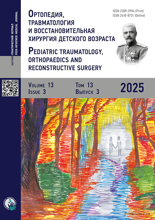
Pediatric Traumatology, Orthopaedics and Reconstructive Surgery

期刊介绍

Pediatric Traumatology, Orthopaedics, and Reconstructive Surgery为季刊,创刊于2013年,是由Turner Scientific Research Institute for Children's Orthopedics of Ministry of Healthcare of Russian Federation 与 Eco-Vector LLC创办的科学学术期刊。

本刊目标读者包括科研人员、内科医师、创伤骨科医师、烧伤医师、小儿外科医师、麻醉医师、儿科医师、神经科医师、口腔外科医师及相关医学领域的所有专科医师。

主编

教授Baindurashvili A.G. (ORCID: 000-0001-8123-6944)

期刊内容:

  • 国内外临床及实验研究成果,外科疾病、烧伤及其所致疾病、肌肉骨骼系统损伤及病变的新型诊治方法研究与资讯;
  • 发表于国际期刊的期刊主题要点、创伤及骨科护理方法与管理文章、案例研究、文献综述、论文摘要;
  • 学术会议与活动的回顾及通知。

索引

  • 本刊被列入“科学学位论文必投的领先同行评期刊目录”。

本刊已被以下国际数据库及各目录收录:

封面Turner Institute for Children's Orthopedics就诊患者的画作

最新一期

开放存取 开放存取  受限制的访问 ##reader.subscriptionAccessGranted##  受限制的访问 订阅或者付费存取

卷 13, 编号 1 (2025)

封面

完整期次

开放存取 开放存取
受限制的访问 ##reader.subscriptionAccessGranted##
受限制的访问 订阅或者付费存取

Clinical studies

Diagnostic value of synovial calprotectin (S100A8/A9) in differential diagnosis of juvenile idiopathic arthritis and pigmented villonodular synovitis in children: preliminary results of a single-center study
Kozhevnikov A., Derkach E., Porohova A., Lukyanov S.
摘要

BACKGROUND: Juvenile idiopathic arthritis is the most common chronic inflammatory musculoskeletal disease in children. Its most prevalent clinical subtype is oligoarthritis. Pigmented villonodular synovitis, also known as tenosynovial giant cell tumor, is a rare benign synovial disorder, which may clinically resemble oligoarthritis. Differential diagnosis between juvenile idiopathic arthritis and pigmented villonodular synovitis is challenging. Intra-articular steroids used in juvenile idiopathic arthritis therapy may induce negative effects in patients with pigmented villonodular synovitis. Orthopedic and surgical procedures used to rule out pigmented villonodular synovitis are burdensome for children. Magnetic resonance imaging may yield similar findings for both conditions at early stages. Several studies revealed that serum calprotectin is a promising biomarker for juvenile idiopathic arthritis.

AIM: To assess synovial fluid calprotectin concentrations in children with oligoarthritis and pigmented villonodular synovitis.

METHODS: The synovial fluid concentrations of calprotectin, interleukin-6, and tumor necrosis factor-alpha in 42 children with oligoarthritis and 12 children with diffuse pigmented villonodular synovitis of the knee joint were obtained by enzyme-linked immunosorbent assay. In patients with juvenile idiopathic arthritis, cytokine levels were determined at disease onset, and prior to therapeutic and diagnostic arthroscopy in those with pigmented villonodular synovitis.

RESULTS: Synovial calprotectin significantly increased in children with oligoarthritis (108 [28.2; 237] μg/mL) compared to those with pigmented villonodular synovitis (1.53 [1.26; 1.69] μg/mL; p < 0.001). No statistically significant differences were found in synovial tumor necrosis factor-alpha and interleukin-6 concentrations between patients with juvenile idiopathic arthritis and those with pigmented villonodular synovitis. ROC analysis showed a synovial calprotectin threshold of >2.9 μg/mL for the diagnosis of juvenile idiopathic arthritis (AUC = 0.996 ± 0.00479; 95% CI: 0.926–1.000).

CONCLUSION: In children, the differential diagnosis of oligoarthritis is often complicated by clinically similar nonrheumatic joint disorders. The main synovial proinflammatory markers cannot be used for the differential diagnosis of juvenile idiopathic arthritis and pigmented villonodular synovitis. Synovial calprotectin concentration is a promising biomarker of juvenile idiopathic arthritis.

Pediatric Traumatology, Orthopaedics and Reconstructive Surgery. 2025;13(1):5-13
pages 5-13 views
Idiopathic avascular necrosis of the femoral head in young rhythmic gymnasts: a clinical and radiological assessment
Pozdnikin I., Bortulev P., Barsukov D., Murashko T.
摘要

BACKGROUND: Osteonecrosis of the femoral head is a severe and rapidly progressive degenerative-dystrophic disorder and is commonly detected in children professionally engaged in rhythmic gymnastics. It is characterized by destructive changes in the femoral head, resulting in hip joint function impairment and, consequently, poor clinical outcomes. The radiographic anatomy of the hip joints is insufficiently studied, especially considering the impact of intense and specific physical loads, course and progression of the disease, and reasons behind its delayed diagnosis and late initiation of treatment.

AIM: To provide a clinical and radiological characterization of the hip joints in young rhythmic gymnasts diagnosed with idiopathic avascular necrosis of the femoral head and identify factors contributing to delayed diagnosis and late treatment initiation.

METHODS: Medical histories and imaging findings of 45 young rhythmic gymnasts aged 12–17 years diagnosed with idiopathic avascular necrosis of the femoral head were analyzed.

RESULTS: Most patients exhibited a severe clinical course due to total or subtotal involvement of the femoral head. Distinctive anatomical features of the hip joint in young rhythmic gymnasts included dysplastic morphology with impaired stability indices, valgus epiphyseal formation resembling a Kalamchi type II deformity, and a tendency toward acetabular retroversion. These characteristics contributed to the development of pathological changes, such as femoral head extrusion and deformation. Delayed diagnosis and initiation of treatment was due to insufficient awareness of this pathology among parents, coaches, and physicians; gradual development of clinical symptoms without a history of overt trauma; and diagnostic errors during the early stages of the disease.

CONCLUSION: In adolescents, their congenital anatomical features of the hip joint contribute to a more severe course of avascular necrosis of the femoral head. Early differential diagnosis of hip pain in gymnasts, including the use of magnetic resonance imaging, is crucial to exclude femoral head osteonecrosis.

Pediatric Traumatology, Orthopaedics and Reconstructive Surgery. 2025;13(1):14-25
pages 14-25 views
Comparative analysis of arthroscopic techniques for fixation of tibial intercondylar eminence avulsion fractures in children
Salikhov M., Avramenko V., Batalov G., Kemkin V.
摘要

BACKGROUND: Avulsion fracture of the tibial intercondylar eminence is a rare injury, which commonly occurs in adolescents aged 8–14 years, and may result in disability caused by improper bone union. Several surgical techniques for the treatment of patients with such fractures have been developed, involving various methods of fixation for the avulsed fragment of the tibial intercondylar eminence.

AIM: To evaluate the effectiveness of the novel technique for the treatment of type III tibial intercondylar eminence avulsion fractures according to the Meyers–McKeever–Zariczny classification in children with open growth plates and compare its outcomes to those of arthroscopically assisted reduction and fixation of the avulsed fragment using a Herbert screw.

METHODS: Functional outcomes in 45 children aged 14–17 years with tibial intercondylar eminence fractures were analyzed at 3, 6, and 12 months after surgery. Group A included 22 children who underwent arthroscopic fixation with a Herbert screw. Group B comprised 23 children who underwent arthroscopic fixation using a self-tightening suture loop following the novel method.

RESULTS: Group B demonstrated better anteroposterior and rotational stability of the knee than group A. Postoperative functional assessment scores (IKDC 2000, Lysholm Knee Scoring Scale, and Tegner Activity Scale) were significantly better in group B than in group A (p = 0.00006). In group A, postoperative complications were observed in 18.1% of cases (p ≤ 0.05), including screw fracture (4.5%) and aseptic synovitis due to a foreign body reaction (13.6%). No postoperative complications were noted in group B (p < 0.05).

CONCLUSION: The proposed technique for treating tibial intercondylar eminence avulsion fractures in children with open growth plates is more reliable and safer compared with Herbert screw fixation and may be recommended for clinical practice.

Pediatric Traumatology, Orthopaedics and Reconstructive Surgery. 2025;13(1):26-37
pages 26-37 views
Inversion injury of ankle joint and foot in children: association with sports participation and comorbidities (analysis based on medical information system data)
Sapogovskiy A., Kenis V., Agranovich O., Trofimova S., Abramov I., Petrova E., Kasev A.
摘要

BACKGROUND: Ankle joint and foot injuries are the most common type of musculoskeletal trauma in children. Their mechanism of injury mainly determines the pattern of damage.

AIM: To optimize the diagnosis and treatment of inversion injuries of the ankle joint and foot in children and assess their association with sports participation and comorbidities.

METHODS: Outpatient medical records of patients treated at the consultative and diagnostic department between 2014 and 2023 were analyzed. Overall, 1518 cases involving ankle joint and foot injuries were determined, including 111 patients referred with a preliminary diagnosis of sprain and an inversion injury mechanism.

RESULTS: The study included boys and girls aged 10–16 years, one-third of them participated in sports, primarily team and gymnastic disciplines. Recurrent inversion injuries occurred more often in children who participated in sports, which should be considered when planning training programs and return to sport. Analysis of the incidence of injuries associated with inversion trauma showed that ligament sprains were most common in nonathlete children (39%), whereas bone fractures predominated in children who participated in sports (38%).

CONCLUSION: This study revealed a high incidence of bone and ligament injuries associated with inversion trauma in children and elucidated the contribution of sports participation to these injuries. The broader use of magnetic resonance imaging and ultrasound, along with the refinement and standardization of their protocols, improves the understanding of inversion injuries of the foot and facilitates development of more effective diagnostic and treatment algorithms.

Pediatric Traumatology, Orthopaedics and Reconstructive Surgery. 2025;13(1):38-48
pages 38-48 views
Differentiated stabilometric assessment of postural control in nonathlete children and comparison with young athletes
Nikityuk I.
摘要

BACKGROUND: Monitoring the functional parameters of the musculoskeletal system is crucial for evaluating children’s adaptive responses to physical activity. Such health monitoring of children participating in sports should be systematic. Stabilometry is a promising method for the functional assessment of the musculoskeletal system in young athletes.

AIM: To develop criteria for the differentiated assessment of postural control in children based on analysis of asymmetry in stabilometric parameters of the contralateral lower limbs centers of pressure and compare these findings with those obtained in young athletes.

METHODS: A two-stage study of postural balance in healthy children was conducted using dual stabilometric platforms. In stage 1, a control group including 59 children aged 5–16 years who were not involved in sports and whose physical activity did not exceed routine daily levels was investigated to determine the range of normal stabilometric parameters. The children were divided into four age subgroups: 16 children aged 5–6 years, 13 aged 7–10 years, and 15 each aged 11–13 years and 14–16 years. In stage 2, 15 boys and girls aged 7–10 years who regularly trained at sports schools in Saint Petersburg were examined.

RESULTS: In the control group, postural balance gradually improved with age starting from 5 to 6 years and reached its highest levels by age 14–16 years. In the main group, regular sports training with increased physical loads in children aged 7–10 years did not adversely affect postural balance or the balance between contralateral lower limbs. This may be associated with a gentle exercise methodology that provides balanced and age-appropriate training loads adjusted for the physiological characteristics of children. The quality of postural control was better in young athletes than in their nonathlete peers, with median stabilometric parameters of 80% and 72%, respectively (p < 0.05). Lower center of pressure velocities and smaller statokinesiogram areas were noted in athletes, showing more efficient postural balance. These findings indicate adaptive processes in the postural ontogenesis of children in the main group because of systematically graded physical loads on the musculoskeletal system.

CONCLUSION: The functional asymmetry between the contralateral lower limbs should be considered, which is a physiological characteristic of growing children, particularly young athletes. This asymmetry is linked to age-related physiological differences in the centers of pressure movements of the left and right lower limbs associated with cerebral lateralization. Considering motor asymmetry of the contralateral lower limbs in training programs is advisable to prevent sports-related injuries.

Pediatric Traumatology, Orthopaedics and Reconstructive Surgery. 2025;13(1):49-61
pages 49-61 views

New technologies in trauma and orthopedic surgery

Patient-specific 3D template with radiopaque markers for pediatric pedicle screw fixation: comparison with freehand technique
Toria V., Vissarionov S., Kubanov R.
摘要

BACKGROUND: Accuracy and precision are crucial in pedicle screw implantation in children with congenital spinal deformities combined with thoracic cage anomalies. The use of patient-specific navigation templates supplemented with radiopaque markers significantly improves screw positioning accuracy and reduces the risk of complications.

AIM: To evaluate the effectiveness of a novel patient-specific surgical template with radiopaque markers and accuracy of pedicle screw placement in children with congenital spinal deformities combined with thoracic cage anomalies, compared with the freehand technique.

METHODS: The study included 26 patients (aged 4–9 years) who underwent surgery at a specialized center. The patients were grouped into two: group 1 (n = 13) underwent screw placement using the new template with radiopaque markers and group 2 (n = 13) underwent screw placement using the freehand technique. Screw placement accuracy was evaluated using the Gertzbein scale based on postoperative radiographs and computed tomography. Statistical analysis was performed using the Student’s t-test, with p < 0.05 indicating significance.

RESULTS: In the navigation template group, 100% of screws were placed correctly (grade 0). In the freehand group, 80.5% of screws were correctly positioned; 16.9% showed deviations up to 2 mm (grade 1), and 2.6% showed deviations >2 mm (grade 2). The use of the navigation template significantly reduced the time required for bone canal preparation compared with the freehand method.

CONCLUSION: Navigation templates with radiopaque markers demonstrate high accuracy and correctness in pedicle screw placement in children with congenital spinal deformities, reducing malposition risk and shortening operative time. Their widespread implementation may enhance the safety and effectiveness of surgical treatment in this patient population.

Pediatric Traumatology, Orthopaedics and Reconstructive Surgery. 2025;13(1):62-69
pages 62-69 views

Exchange of experience

Hip arthroplasty for bilateral pathologic hip dislocation in children
Garkavenko Y.
摘要

BACKGROUND: Pathologic hip dislocation is a severe complication of septic arthritis and hematogenous osteomyelitis of the proximal femoral metaphysis in young children. Bilateral involvement of the hip joints results in more severe anatomical and functional impairments, requiring greater treatment efforts. In such cases, rehabilitation may take years and involves staged surgical correction and continuous conservative management. Meeting these requirements is crucial, as insufficient or inadequate treatment may result in contractures, restricted hip mobility, and progression of secondary deformities.

AIM: To analyze the outcomes of surgical treatment in children with bilateral pathologic hip dislocations treated with hip arthroplasty using demineralized osteochondral allografts and highlight rehabilitation aspects required to achieve favorable short- and long-term postoperative outcomes.

METHODS: Surgical outcomes in 11 children (22 hip joints) aged 1.5–10 years with bilateral destructive pathologic hip dislocations who underwent hip arthroplasty using demineralized osteochondral allografts were analyzed.

RESULTS: Long-term follow-up (1–12 years) demonstrated radiologically confirmed joint stability in 86.4% of cases and preservation of at least 80 of hip flexion range.

CONCLUSION: Hip arthroplasty with demineralized osteochondral allografts in children with destructive pathologic hip dislocations is an effective method for restoring hip joint function, providing stability and preserving range of motion over the long term. A consistent, systematic, and comprehensive treatment approach is critical for successful rehabilitation and for restoration and long-term preservation of hip function in children with bilateral hip involvement.

Pediatric Traumatology, Orthopaedics and Reconstructive Surgery. 2025;13(1):70-76
pages 70-76 views

Clinical cases

Surgical treatment of severe pectus excavatum in an adolescent: a case report
Ryzhikov D., Dolgiev B., Vissarionov S., Zhukova J., Boroznyak I.
摘要

BACKGROUND: Pectus excavatum is a malformation characterized by deformities of the cartilaginous parts of the ribs and sternum of varying severity and accounts for 75%–91% of all chest wall deformities. Surgical treatment of pectus excavatum in children remains an urgent issue, despite the considerable number of existing surgical correction techniques. These techniques are not universally applicable, especially for asymmetric and rigid forms, requiring ongoing modification and improvement.

CASE DESCRIPTION: A 17-year-old patient underwent surgical correction of a severe pectus excavatum complicated by a history of cardiac surgery, namely, median sternotomy and mitral valve prosthesis placement. The surgical procedure involved the release of major vital anatomical structures, the use of an external fixation device providing high corrective force during surgery, and gradual intraoperative correction under cardiac function monitoring.

DISCUSSION: Correction of severe pectus excavatum following prior median sternotomy is associated with a high risk of complications, including fatal events such as asystole or massive hemorrhage. In such cases, a minimally invasive thoracoplasty with an additional subxiphoid approach and sternal elevation using an external fixation device is recommended. Conventional sternal elevation techniques (e.g., bone hooks, sutures, and clamps) have been found to be not advisable for pronounced rigid deformities owing to the risk of sternal injury. Moreover, isolated thoracoscopy using the standard MIRPE technique does not ensure the integrity of intrathoracic structures, and traditional bar-flipping maneuver for immediate and forced correction of pectus excavatum is inappropriate.

CONCLUSION: Patients with pectus excavatum exceeding the threshold for severe deformity, particularly those with previous cardiac surgery, require a treatment different from standard thoracoplasty. The presented approach, involving the release of major vital anatomical structures and gradual intraoperative correction under cardiac monitoring with an external fixation device, is recommended for managing such complex clinical cases.

Pediatric Traumatology, Orthopaedics and Reconstructive Surgery. 2025;13(1):77-85
pages 77-85 views
Combined reconstruction of femoral condylar osteochondral defects in adolescents: clinical cases and a review
Semenov S., Zorin V.
摘要
Pediatric Traumatology, Orthopaedics and Reconstructive Surgery. 2025;13(1):86-96
pages 86-96 views

Scientific reviews

Role of genetic determinants in the development of congenital scoliosis: a review
Vissarionov S., Pershina P., Khalchitsky S., Asadulaev M.
摘要

BACKGROUND: Congenital scoliosis is a multifactorial disorder resulting from disturbances during vertebral embryogenesis. Defects occurring at any stage of fetal development may lead to congenital scoliosis and progressive spinal deformity. Recent studies highlighted genetic factors as crucial determinants of the development of this condition.

AIM: To review the sources on the genetic basis of congenital scoliosis, focusing on molecular regulatory mechanisms, mutation frequencies, and the contribution of specific genes.

METHODS: Sources were retrieved using keyword searches in PubMed, Google Scholar, Cochrane Library, Web of Science, Lens.org, and eLibrary over the past 25 years. The inclusion criteria were full-text availability, meta-analyses, systematic reviews, cohort studies of patients with congenital scoliosis, experimental animal models, and case–control studies. The exclusion criteria included lack of full-text access, patents, utility models, and studies without clinical data. Finally, 54 publications were selected for detailed analysis.

RESULTS: The identified genes were categorized into four: susceptibility genes (LMX1A, PTK7, SOX9, TBX6, and TBXT); genes wherein mutations directly cause syndromes or monogenic disorders associated with scoliosis (FBN1); genes with copy number variations (DHX40, DSCAM, MYSM1, and NOTCH2); and genes showing abnormal methylation in patients with scoliosis (COL5A1, GRID1, GSE1, RGS3, SORCS2, IGH1, IGH3, IGHM, KAT6B, and TNS3).

CONCLUSION: Analysis of scientific sources revealed the presence of predisposing genetic factors associated with the development of congenital scoliosis in its various phenotypic forms. Data from large-scale studies clarified the etiological factors and were beneficial for predicting the course of congenital scoliosis, and findings from studies with smaller samples may help define future directions for identifying genetic determinants of this disorder.

Pediatric Traumatology, Orthopaedics and Reconstructive Surgery. 2025;13(1):97-107
pages 97-107 views
Hallux valgus in equino-planovalgus foot deformity in children with cerebral palsy and its etiopathogenesis: a review (part 1)
Umnov V., Zharkov D., Novikov V., Umnov D.
摘要

BACKGROUND: Hallux valgus in children with cerebral palsy is an understudied problem. Treatment approaches are generally applied as a secondary measure, often after the child starts complaining at an older age following correction of contractures and other foot deformities. Moreover, there are no established methods for the early prevention or treatment of hallux valgus. Understanding the fundamental mechanisms of etiopathogenesis and biomechanical disturbances during gait is crucial for developing preventive and therapeutic strategies for this patient population.

AIM: To analyze international studies of foot deformities in children with cerebral palsy and compare these findings with biomechanical studies in patients with idiopathic hallux valgus without neurological pathology.

METHODS: Sixty-four scientific articles and publications retrieved from multiple databases without time restrictions were reviewed.

RESULTS: Equinoplanovalgus foot deformity is a major etiopathogenetic factor in the development of hallux valgus in children with cerebral palsy. Biomechanical alterations associated with hallux valgus are characterized by limited dorsiflexion of the hallux, excessive dorsiflexion of the first ray, restricted supination of the hindfoot and midfoot, and increased plantar flexion of the ankle joint during the terminal stance phase. In equinoplanovalgus deformity, excessive pronation of the hindfoot and midfoot cannot be compensated because of the limited range of motion of the midtarsal joint, causing restricted midfoot supination and the inability to activate the locking mechanisms of the midfoot and forefoot during terminal stance.

CONCLUSION: Any biomechanical disturbance within the complex multisegmental structure of the lower extremity that reduces hindfoot and midfoot supination, causes first ray eversion, and limits hallux dorsiflexion may contribute to deformity. The diversity of motor disorders, contracture patterns, and deformities in children with cerebral palsy indicates the need for further research aimed at identifying the specific factors involved in hallux valgus formation. Such findings may be beneficial for developing preventive and therapeutic strategies for early-stage deformities.

Pediatric Traumatology, Orthopaedics and Reconstructive Surgery. 2025;13(1):108-117
pages 108-117 views

Anniversray

Alla Vladimirovna Ovechkina
摘要

Alla Vladimirovna Ovechkina, Candidate of Sciences in Medicine, Assistant Professor, leading expert at the H. Turner National Medical Research Center for Сhildren’s Orthopedics and Trauma Surgery of the Ministry of Health of the Russian Federation, and Honored Doctor of the Russian Federation, celebrates her 80th birthday. Alla Ovechkina has devoted over 50 years of her professional life to the H. Turner National Medical Research Center for Сhildren’s Orthopedics and Trauma Surgery. In 1988–2025, she served as the center’s Scientific Secretary. She became an indispensable assistant to eight successive directors of the H. Turner Center with her energy, dedication, and impeccable professionalism. Alla Ovechkina’s scientific work has been focused on the conservative management of spinal disorders in children.

Pediatric Traumatology, Orthopaedics and Reconstructive Surgery. 2025;13(1):118-119
pages 118-119 views