Pediatric Traumatology, Orthopaedics and Reconstructive Surgery
期刊介绍
Pediatric Traumatology, Orthopaedics, and Reconstructive Surgery为季刊,创刊于2013年,是由Turner Scientific Research Institute for Children's Orthopedics of Ministry of Healthcare of Russian Federation 与 Eco-Vector LLC创办的科学学术期刊。
本刊目标读者包括科研人员、内科医师、创伤骨科医师、烧伤医师、小儿外科医师、麻醉医师、儿科医师、神经科医师、口腔外科医师及相关医学领域的所有专科医师。
主编
教授Baindurashvili A.G. (ORCID: 000-0001-8123-6944)
期刊内容:
- 国内外临床及实验研究成果,外科疾病、烧伤及其所致疾病、肌肉骨骼系统损伤及病变的新型诊治方法研究与资讯;
- 发表于国际期刊的期刊主题要点、创伤及骨科护理方法与管理文章、案例研究、文献综述、论文摘要;
- 学术会议与活动的回顾及通知。
索引
- 本刊被列入“科学学位论文必投的领先同行评议期刊目录”。
本刊已被以下国际数据库及各目录收录:
- 俄罗斯科学引文索引(Russian Science Citation Index)
- SCOPUS
- Embase
- EBSCO
- DOAJ
- Google Scholar
- Ulrich's Periodical Directory
- WorldCat
- CNKI
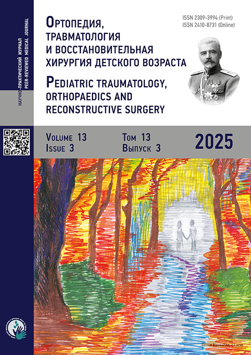
封面:Turner Institute for Children's Orthopedics就诊患者的画作
最新一期


卷 13, 编号 2 (2025)
- 年: 2025
- ##issue.datePublished##: 10.07.2025
- 文章: 11
- URL: https://journals.eco-vector.com/turner/issue/view/13008
- DOI: https://doi.org/10.17816/PTORS.132
Clinical studies
Assessment of sagittal balance parameters in adolescents with severe thoracic idiopathic scoliosis
摘要
BACKGROUND: In recent years, the concept of sagittal balance has become increasingly important in the treatment of spinal deformities of various etiologies. However, the number of studies analyzing sagittal balance parameters in patients with idiopathic scoliosis is limited, especially those involving individuals with severe deformities.
AIM: This study aimed to evaluate global and regional sagittal balance parameters in adolescents with severe thoracic idiopathic scoliosis.
METHODS: The study included 45 patients aged 12–17 years diagnosed with thoracic idiopathic scoliosis (Lenke types 1, 2, and 3), presenting with a primary curve magnitude ranging from 80° to 150°, according to the Cobb method. Radiographic examinations were performed to assess both regional and global sagittal balance parameters, as well as characteristics of frontal plane spinal deformity. The patients were divided into two groups based on the global sagittal balance values. Descriptive, correlation, regression, and comparative statistical analyses of the data were conducted.
RESULTS: A positive correlation was observed between the magnitude of frontal plane spinal deformity and thoracic kyphosis values. The regression analysis indicated that when the primary curve exceeded approximately 100°, thoracic kyphosis values tended to rise above 60°. Thoracic hyperkyphosis was the predominant sagittal deformity in these patients; however, neither thoracic kyphosis nor the primary curve magnitude in the frontal plane showed statistically significant correlation with any of the global sagittal balance indicators. These indicators correlated more strongly with spinopelvic parameters (pelvic incidence (PI), pelvic tilt (PT), sacral slope (SS), lumbar lordosis (LL), and PI–LL).
CONCLUSION: Surgical treatment of severe idiopathic scoliosis should obligatorily consider correction methods that produce a corrective effect on the kyphotic component of the deformity. The development of pronounced anterior imbalance has been proven to be uncharacteristic for patients with thoracic idiopathic scoliosis, even with the largest primary curve magnitudes. The primary risk factors for its development were reduced PI and LL values. The development mechanism of the observed association in these patients remains unclear.


Transitional lumbosacral vertebrae in children with pelvic fractures: frequency of diagnosis and distribution of types and subtypes
摘要
BACKGROUND: Various aspects of transitional lumbosacral vertebrae in children remain relevant due to a lack of research on several critical issues. For instance, the frequency of diagnosis and the distribution of different types and subtypes of this condition in the pediatric population remain unknown. The clinical features, particularly pain as the main symptom, have not been sufficiently studied. Furthermore, effective and pathogenetically based approaches to the treatment and prevention of low back pain associated with transitional lumbosacral vertebrae in different pediatric groups have not yet been developed.
AIM: The study aimed to determine the prevalence and structure of transitional lumbosacral vertebrae in children with pelvic fractures.
METHODS: This study included 41 children who sustained pelvic fractures between 2022 and 2024, with transitional lumbosacral vertebrae in 10 patients. The diagnostic protocol adhered to the standard for patients with pediatric trauma and included mandatory computed tomography of the lumbar spine and pelvis. Pelvic fractures were classified according to the Tile/AO classification system. Acetabular fractures were assessed using the classification criteria proposed by Judet et al. Transitional lumbosacral vertebrae types and subtypes were categorized based on the Castellvi classification.
RESULTS: The diagnostic frequency of transitional lumbosacral vertebrae was found to be 24.4% ± 6.7% of clinical cases. Subtype IIa was the most common, accounting for 50.0% ± 15.8% of cases, followed by subtype IIIb, which occurred in 30.0% ± 14.5% of patients. Subtypes Ia and IIb each represented 10.0% ± 9.4% of the observed cases. The study revealed that, unlike in adult patients, a distinguishing feature of the condition in children was the absence of the main symptom, namely, pain in the lumbosacral junction.
CONCLUSION: The high diagnostic frequency of this condition, which often remains latent for some time, highlights the importance of targeted radiological assessment of the lumbosacral junction. Once transitional lumbosacral vertebrae are identified, patients should be informed of their presence to support the joint development of individualized strategies for preventing lumbosacral pain.


Clinical and MRI features of patellar instability in children
摘要
BACKGROUND: Patellar instability refers to recurrent dislocations and subluxations of the patella relative to the femoral trochlear groove. This condition is one of the most common disorders of the knee joint in pediatric patients. Both bone and soft tissue structures act as stabilizers of the patella, and alterations in any of these components may contribute to patella instability. Magnetic resonance imaging (MRI) findings related to the osteochondral and soft tissue structures of the patellofemoral joint in children with patellar instability, as well as their correlation with clinical presentation, are of practical interest.
AIM: This study aimed to assess the key MRI features of the patellofemoral joint zone in pediatric patients with patellar instability and evaluate their clinical manifestations.
METHODS: Epidemiological, clinical, and MRI data were analyzed for patients with patellar instability and anterior cruciate ligament injury. The study included 52 patients in the main group and 44 patients in the comparison group with anterior cruciate ligament injury. No statistically significant differences in age distribution were observed.
RESULTS: Significant differences were identified between the groups in terms of Wiberg patellar type, presence of a patellar apprehension sign, and patellar hypermobility. Differences were also noted in the lateral inclination and depth of the trochlear groove, as well as in the frequency of clinical signs such as patellar apprehension and patellar hypermobility (p < 0.001).
CONCLUSION: Trochlear dysplasia is a key predisposing factor for the development of patellar instability in pediatric patients. This study confirmed statistically significant differences in the parameters characterizing trochlear dysplasia, as well as the influence of this factor on patellar instability in children, including its correlation with clinical manifestations.


A Photometric Method for Measuring Ankle Dorsiflexion Reproducibility in Young Athletes
摘要
BACKGROUND: Ankle dorsiflexion is one of the most important biomechanical indicators of the potential functional capacity of the foot. Its quantitative assessment is crucial in clinical evaluation, and in determining children’s readiness for sports and their potential risk of sports-related injuries.
AIM: This study aimed to evaluate the reproducibility of a photometric method for measuring ankle dorsiflexion angles in young athletes in relation to the conventional goniometric method.
METHODS: This study included 30 children aged 6–12 years who participated in regular sports activities and had not been diagnosed with orthopedic disorders. Dorsiflexion was measured by two independent specialists using goniometry and photometry based on anatomical landmarks. Inter-rater reliability was assessed using the intraclass correlation coefficient (ICC). The agreement between the methods was analyzed using Bland–Altman plots.
RESULTS: There were statistically significant differences (t-test: p = 0.04) in mean dorsiflexion angles between the goniometry measurement (17.15°) and the photometry measurement (18.1°). Photometry-based measurements exhibited higher inter-rater reliability (ICC = 0.91) than goniometry-based measurements (ICC = 0.78).
CONCLUSION: These findings indicate that the photometric method of measuring ankle dorsiflexion in children is a reliable and reproducible tool that can be recommended for practical use and monitoring for physically active children.


Organization of trauma and orthopedic care
Prevalence of Musculoskeletal Diseases Among Children in Saint Petersburg, Russia
摘要
BACKGROUND: Most musculoskeletal diseases are diagnosed in childhood, allowing for the prevention of chronic progression and subsequent disability in adulthood. In recent years, monitoring pediatric morbidity has gained increasing scientific and practical importance, leading to the reassessment of key organizational strategies.
AIM: This study aimed to conduct a comparative analysis of the overall morbidity of musculoskeletal and connective tissue diseases among children in Saint Petersburg from 2017 to 2023.
METHODS: Based on official statistical reports from 2017 to 2023, the prevalence of musculoskeletal and connective tissue diseases among children aged 0–14 years and adolescents aged 15–17 years in Saint Petersburg was compared with national averages in Russia. Morbidity trends were analyzed per 1,000 of the respective pediatric population. The analysis was carried out using a quantitative assessment of the odds ratio for the occurrence of musculoskeletal conditions, overall and by individual disease forms, as presented in the statistical bulletins. Statistical significance was inferred when the 95% confidence interval for the odds ratio did not include 1 (p < 0.05). Data analysis was performed using Microsoft Office 2010 (Word and Excel).
RESULTS: The prevalence of musculoskeletal and connective tissue disorders among children in Saint Petersburg was extremely high, showing an upward trend and a widening gap compared with the national average in Russia. During the COVID-19 pandemic, the growth rates of most disease categories outpaced those of the preceding period. Among children aged 0–14 years in Saint Petersburg, the odds ratio showed an increase for reactive arthropathies (by 23.6%), deforming dorsopathies (by 27.4%), and other musculoskeletal conditions (by 23.0%). Among adolescents, increased odds were observed for juvenile arthritis (7.9%), arthropathies overall (13.3%), deforming dorsopathies (15.5%), and other disorders (22.7%). In contrast, both age groups showed a decreased likelihood of being diagnosed with spondylopathies. When assessing the significance of odds ratios and the validity of conclusions, preference should be given to the use of the smallest measurement units.
CONCLUSION: An increase in the prevalence and odds of musculoskeletal and connective tissue disorders among children in Saint Petersburg was observed. This underscores the need to improve care pathways and adopt modern approaches to the prevention and treatment of these conditions in the region, as well as to strengthen the availability and quality of medical resources in other Russian regions.


New technologies in trauma and orthopedic surgery
Assessment of amplitude-frequency characteristics of postural sway at the pressure centers of the contralateral lower limbs of young athletes
摘要
BACKGROUND: The development of instrument-based methods for the functional diagnostics of adaptive body changes in children engaged in sports activities is a currently relevant area of research.
AIM: This work examined the amplitude characteristics of postural sway at the pressure centers of the contralateral lower limbs in young athletes in motionless standing positions across a frequency range.
METHODS: Amplitude-frequency characteristics were analyzed during motionless standing in 18 children aged 7–10 years, of both sexes, who regularly participated in track and field. The postural sway parameters of the pressure centers under both feet were recorded using two stabilometric platforms. Measurements were collected in the low-frequency range (up to 1.0 Hz) for 20 seconds and the high-frequency range (1.0–6.0 Hz) for 1 second. The deviation angle α (°) of the sway axis of the lower limbs from the midline axis was determined. The first peak amplitudes and frequencies of sway were measured: а1, а2, and а3 (mm) and f1, f2, and f3 (Hz) in the low-frequency range, respectively; and А1, А2, and А3 (mm) and F1, F2, and F3 (Hz) in the high-frequency range. The control group comprised 18 children with typical daily physical activity levels.
RESULTS: In young athletes, more favorable amplitude-frequency characteristics of postural sway at the low- and high-frequency ranges were identified compared with age-matched peers not engaged in sports and leading sedentary lifestyles. This effect was manifested by a frequency shift in sway, a reduction in all peak sway amplitudes at both the low- and high-frequency ranges, lower asymmetry of sway amplitudes, and the stable synchronization of pressure center oscillations across the entire frequency spectrum.
CONCLUSION: Amplitude-frequency parameters of postural sway at the pressure centers of the contralateral lower limbs should be considered while training younger school-aged children for sports to prevent neuromuscular imbalance due to fatigue, and injuries.


Clinical cases
Ischemic stroke of the cervical spinal cord: a review and case report
摘要
BACKGROUND: Ischemic spinal cord stroke is a rare condition, accounting for approximately 1% of all spinal cord strokes. The relevance of this publication is determined by the rarity of the condition, the complexity of differential diagnosis with other acute onset myelopathic syndromes, the severity of spinal stroke outcomes, and insufficient awareness among physicians regarding this condition in children.
CASE DESCRIPTION: We present a clinical case of acute myelopathic syndrome in an 8-year-old child. Magnetic resonance imaging of the cervical spinal cord correlated with the clinical findings and indicated impaired circulation in anterior spinal artery at the cervical spinal level.
DISCUSSION: Acute impairment of spinal cord circulation may be caused by systemic hypotension; occlusion of spinal cord-supplying vessels (traumatic, iatrogenic, thrombotic, or embolic); arterial dissection; arteriovenous malformations and fistulas; or hypercoagulable states.
CONCLUSION: In pediatric patients presenting with acute myelopathic syndrome, ischemic stroke of the spinal cord should be considered in the differential diagnosis alongside inflammatory and infectious conditions, particularly in children with significant risk factors. Early recognition of acute impairment of spinal cord circulation is particularly important for timely neuroimaging, consultations with related specialists, and initiating etiotropic or symptomatic treatment upon identification of the underlying cause of acute spinal cord ischemia, as well as early rehabilitation.


Scientific reviews
Enchondromas of the hand in children: current concepts in etiopathogenesis, diagnosis, and treatment: a review
摘要
The prevalence of benign bone tumors among pediatric patients highlights the relevance of such investigations for orthopedic practice. This article focuses on hand enchondromas in children. Despite the relative frequency of this condition, the lack of clinical guidelines for this disease area underscores the relevance of the publication. A total of 69 full-text articles were selected from 387 sources identified through searches in CyberLeninka, eLibrary, PubMed, and Scopus from 1990 to 2023. This review presents current views on the etiopathogenesis, diagnostic and therapeutic challenges, and prognosis of hand enchondromas in children. Radiography and computed tomography are the primary diagnostic methods for hand enchondromas. Histological verification of diagnosis is mandatory. Particular attention should be given to surgical treatment strategies, including the optimal timing and extent of intervention, selection of grafting material, risk factors, and frequency of recurrence. The scientific data analysis enabled the systematization of data on the etiopathogenesis of hand enchondromas, identification of diagnostic priorities, clarification of diagnostic and differential diagnostic criteria, and refinement of approaches to treatment strategy and methods. The choice of treatment strategy, including the method and timing of intervention, should be individualized, taking into account tumor localization and histogenesis, as well as any additional diagnostic findings and the risk of complications. In recent years, specialists have tended to favor early surgical intervention over wait-and-see approaches, with the choice between autograft and allograft being determined by the clinical context. Autografts are considered the gold standard; however, the use of allografts for restoring the structural and functional integrity of bone in cases of injury is a reasonable and well-substantiated alternative, even in the light of the high risks of donor site morbidity. Such decisions should be made collaboratively, considering the opinions of the involved specialists, including orthopedic oncologists, radiologists, pathologists, and rehabilitation physicians.


Surgical treatment of knee flexion contracture in patients with cerebral palsy: a review
摘要
Knee flexion contracture is the most common lower limb deformity among patients with cerebral palsy, with a prevalence of 47%–53%. Its prolonged persistence leads to marked functional impairments, such as increased energy expenditure during walking and decreased motor activity in children, and the emergence of secondary orthopedic complications, which significantly affects patients’ quality of life. This review analyzed published scientific data on current surgical methods for the correction of knee flexion contracture in patients with cerebral palsy, evaluating their clinical effectiveness, determining frequency of complications, and identifying factors influencing the choice of optimal treatment strategy. The studies were determined using PubMed, Google Scholar, Cochrane Library, Crossref, and eLibrary. The search was conducted without language restrictions, covering the period from 1952 to 2024. Seventy-four sources were reviewed, including original articles and systematic reviews on surgical treatment of knee flexion contracture in patients with cerebral palsy. The primary surgical correction methods for knee flexion contracture in children with cerebral palsy are hamstring lengthening and distal femoral extension supracondylar osteotomy. Hamstring lengthening is considered the main surgical procedure and most appropriate option for patients of all age groups. Notably, femoral osteotomy has shown high clinical effectiveness in older patients, particularly in those with marked distal femoral deformities. Both methods yield favorable outcomes, but may be associated with certain complications. Furthermore, this review discusses alternative techniques, including tendon transfers and temporary hemiepiphysiodesis, and combined approaches. The conducted scientific data review confirmed the effectiveness of surgical treatment of knee flexion contracture in patients with cerebral palsy. However, in selecting a particular surgical procedure, individual anatomical and clinical features, the degree of contracture, and age-related patient characteristics should be considered.


Stress fractures in upper extremities of athletes: a review
摘要
Stress (overuse) fractures of the upper extremities are a relatively uncommon condition typically observed in athletes. Due to their gradual onset, typically mild clinical symptoms, and sometimes unremarkable imaging findings, such injuries are frequently overlooked. The article aims to present data on the pathogenesis, clinical presentation, diagnostics, and treatment strategies of COVID-19. A scientific data search was conducted in the PubMed (MEDLINE) database and via the Google search engine from 1975 to 2024 using combinations of the operators OR and the following keywords: стрессовые переломы (stress fractures), нагрузочные переломы (overuse fractures), верхние конечности (upper extremities), and спортсмены (athletes). A total of 70 sources were selected, including 22 published within the past decade. Stress (overuse) fractures are reported in approximately 40% of athletes, with 80%–95% occurring in the lower extremities and only 5%–20% occurring in the upper extremities. Stress fractures result from repetitive mechanical loading of the bone and are compounded by fatigue of the surrounding muscles, leading to an increase in the rate of bone resorption by osteoclasts to exceed the rate of new bone formation by osteoblasts. Magnetic resonance imaging is the primary diagnostic modality. In most cases, bone healing occurs with load restriction and activity modification (avoidance of repetitive trauma-inducing movements). Surgical intervention is often required in stage III gymnast wrist fractures, avulsion fractures of the medial epicondyle with displacement ≥10 mm, complete or partial tears of the ulnar collateral ligament of the elbow, olecranon fractures, and carpal bone fractures. Although relatively rare, upper extremity stress fractures can cause significant problems and hinder athletic progression. A thorough medical history, physical examination, and analysis of the mechanism of injury will allow clinicians to raise suspicion of this condition. Load restriction, elimination of provoking factors, gradual increase in training intensity, and improvement of sports technique, in most cases, allowed the patient to avoid surgical treatment and return to sports activities.


Obituaries
Eduard V. Ulrikh (1937–2025)