Pediatric Traumatology, Orthopaedics and Reconstructive Surgery
Scientific academic journal published four times a year since 2013.
- Since 2016 the journal publishes articles in Russian and English in parallel
- Since 2018 in Chineze in additional
- Special issues (conference proceedings) are published in Russian.
Founders
Publisher
Editor-in-Chief
- Baindurashvili A.G., MD, PhD, Professor
ORCID: 000-0001-8123-6944
About
The target audience of the journal is researches, physicians, orthopedic trauma, burn, and pediatric surgeons, anesthesiologists, pediatricians, neurologists, oral surgeons, and all specialists in related fields of medicine.
The journal publishes original articles:
- results of clinical and experimental research with new data on diagnostic and treatment for patients with surgical diseases, burns and their consequences, injuries and disorders of the musculoskeletal system;
- lecture notes on journal topics, guidelines articles on organization (and management) of trauma and orthopaedic care, case reports, reviews and short communications;
- editorials and news for health care professionals in the appropriate field of medicine .
Indexation
- Russian Science Citation Index
- SCOPUS
- Embase
- Cyberleninka
- Google Scholar
- Ulrich's Periodical Directory
- Dimensions
- DOAJ
- CNKI
- Publons
Types of manuscripts to be accepted for publication
- systematic reviews
- results of original research
- clinical cases and series of clinical cases
- experimental work (technical development)
- datasets
- letters to the editor
Publications
- quarterly, 4 issues per year
- continuously in Online First (Ahead of Print)
- in English, Russian and Chineese (full-text translation)
- with Green Open Access and Optional Gold Open Access for authors
Distribution
- in hybrid mode: by subscription and in Open Access (under the CC BY-NC-ND 4.0)
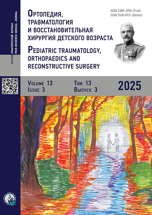
On the cover – drawing of the patient from the H.Turner National Center for Children’s Orthopedics.
Current Issue
Vol 13, No 3 (2025)
- Year: 2025
- Published: 30.10.2025
- Articles: 12
- URL: https://journals.eco-vector.com/turner/issue/view/13746
- DOI: https://doi.org/10.17816/PTORS.133
Clinical studies
Comparative Analysis of Surgical Treatment Outcomes in Children With Unstable Distal Radius Fractures
Abstract
BACKGROUND: Distal radius fractures are common injuries of the musculoskeletal system among children. Poor treatment outcomes in this group of patients require repeated interventions and are associated with limited functional activity of the upper limb. Therefore, the development of new surgical techniques for the treatment of unstable distal radius fractures in children, which minimize the risk of complications and improve rehabilitation outcomes, remains highly relevant.
AIM: This study aimed to compare the surgical outcomes in children with unstable distal radius fractures treated using two different techniques.
METHODS: Surgical treatment was performed in 83 children with unstable distal radius fractures. Group 1 underwent surgery using modified antegrade intramedullary osteosynthesis with a pre-bent Kirschner wire (patent No. 2835501; January 20, 2025) (n = 52). Conversely, group 2 was treated using retrograde intramedullary osteosynthesis with a Kirschner wire (n = 31). Outcomes were assessed at 1, 3, 6, and 12 months by evaluating postoperative complications, contractures of adjacent joints, operative time, and fracture healing rates. Statistical analysis was conducted using the Mann–Whitney test, Yates-corrected χ2 test, and Fisher’s exact test.
RESULTS: The patients treated with retrograde intramedullary osteosynthesis with a Kirschner wire showed a higher incidence and severity of postoperative complications, with less favorable outcomes, compared with those who underwent the modified antegrade osteosynthesis technique. The proposed method did not present greater procedural complexity than did the classical approach, as confirmed by the absence of significant differences in operative time. Long-term functional outcomes were superior in children treated with the modified technique. This indicates that this approach is not only competitive but also preferable.
CONCLUSION: This study supports the use of the proposed modified technique for the surgical treatment of children with unstable distal radius fractures, with the aim of decreasing postoperative complication risk and improving rehabilitation outcomes.
 237-246


Analysis of Surgical Techniques for Avulsion Fractures of the Distal Phalanges in Children
Abstract
BACKGROUND: Avulsion intra-articular fractures account for up to 18% of all bony injuries of the distal phalanx among children. Currently, there is no unified approach for the surgical treatment of such injuries. The Ishiguro technique is an alternative to open reduction; however, experience with its use in pediatric traumatology is limited.
AIM: This study aimed to determine the optimal surgical technique for avulsion intra-articular fractures of the distal phalanges in children by comparing the outcomes of open reduction with fixation of the peripheral fragment and of minimally invasive reduction with osteosynthesis using the Ishiguro method.
METHODS: A prospective cohort study was conducted at Traumatology Departments Nos. 1 and 2 of the Children’s City Clinical Hospital No. 9, Yekaterinburg. Twenty-nine children with avulsion intra-articular fractures of the distal phalanges, with displacement greater than one-third of the articular surface, were included. In the main group (n = 15), minimally invasive reduction and osteosynthesis using the Ishiguro technique were performed; in the control group (n = 14), open reduction and fixation of the peripheral fragment with a wire were conducted. Local inflammatory changes were assessed on postoperative days 3 and 7. The range of motion in the distal interphalangeal joint was measured using a goniometer at 1, 2, and 4 weeks after implant removal.
RESULTS: The main group demonstrated significantly more effective restoration of distal interphalangeal joint motion: 17.80° ± 7.43° versus 7.79° ± 3.40° at 1 week after implant removal (p < 0.001); 57.47° ± 13.11° versus 28.86° ± 12.09° at 2 weeks (p < 0.001); and 89 [85; 90]° versus 78.50 [77.25; 83.00]° at 4 weeks (p < 0.001). Macroscopic evaluation of pin entry sites on postoperative days 3 and 7 showed no significant differences between the groups (p > 0.05). Fracture consolidation was achieved in all cases. Peripheral fragment comminution was observed in one patient from the control group.
CONCLUSION: The Ishiguro osteosynthesis technique may be recommended as the method of choice for treating children with intra-articular avulsion fractures of the distal phalanges owing to its technical reproducibility, minimal invasiveness, and favorable functional outcomes.
 247-255


Correction of Knee Flexion Contracture in Children With Cerebral Palsy by Femoral Extension Osteotomy: Evaluation of the Sagittal Profile
Abstract
BACKGROUND: Knee flexion contracture is one of the most common deformities in children with cerebral palsy, significantly affecting the patients’ gait, energy expenditure, verticalization, and quality of life. Knee flexion contracture can be corrected using either soft tissue procedures (hamstring lengthening) or bony interventions (femoral extension osteotomies). Soft tissue procedures are considered less invasive and are justified based on the underlying pathology. Some studies have reported their effect on sagittal balance, specifically an increase in anterior pelvic tilt. Femoral extension osteotomies have been regarded as sagittally neutral; however, most studies included them as part of combined interventions, which precludes assessment of the isolated effect of the osteotomy itself. This emphasizes the importance of investigating the impact of femoral extension osteotomies on global sagittal alignment in children with cerebral palsy.
AIM: This study aimed to evaluate the effect of corrective femoral extension osteotomy on sagittal spinopelvic parameters in children with cerebral palsy and knee flexion contracture.
METHODS: The study included 14 patients with cerebral palsy treated at the Turner National Medical Research Center for Children’s Orthopedics between 2022 and 2025. The patients underwent corrective supracondylar femoral extension osteotomy with plate fixation with angular stability (LCP PHP 90°). Overall, 26 osteotomies were performed. In three cases, a newly developed implant designed for patients with cerebral palsy with reduced bone density was used. Clinical outcomes (i.e., active extension deficit, contracture degree, and popliteal angle) and radiological parameters (i.e., pelvic incidence, pelvic tilt, sacral slope, lumbar lordosis, thoracic kyphosis, and sagittal vertical axis) were assessed preoperatively and at 6 months postoperatively.
RESULTS: Significant correction of contracture and improvement in active knee extension were observed. Among the radiological parameters, only lumbar lordosis showed a significant change (+4.3° ± 13.5°, p = 0.049). Other parameters remained unchanged. No associations were found between the changes in clinical and radiological parameters.
CONCLUSION: Femoral extension osteotomy is an effective method for correcting knee flexion contracture in children with cerebral palsy and does not cause global sagittal alignment disruption. The increase in lumbar lordosis is adaptive in nature and is not associated with signs of decompensation. Initial experience with the newly designed implant demonstrated technical reliability of fixation and promising applicability in patients with reduced bone density.
 256-265


Ultrasound Criteria of Reparative Osteogenesis in the Distraction Regenerate of the Femur in Patients Aged 9–12 Years With Achondroplasia Undergoing Cross-Lengthening of the Femur and Contralateral Tibia
Abstract
BACKGROUND: The relevance of longitudinal assessment of reparative osteogenesis is shown by the fact that the maturation of the femoral regenerate defines the timing of initiating contralateral tibial lengthening and overall hospital stay duration.
AIM: This study aimed to identify ultrasound criteria of reparative osteogenesis in the femoral distraction regenerate that allow for the initiation of surgical treatment on the contralateral tibia in patients with achondroplasia.
METHODS: Patients with achondroplasia aged 9–12 years (n = 37) were examined; at age 6–7 years, they had undergone the first stage of treatment, namely, bilateral tibial lengthening. At the second stage of treatment, femoral lengthening was performed first, followed by contralateral tibial lengthening. Femoral lengthening was achieved using double corticotomy. The distraction period lasted 63 ± 3 days, with a length gain of 6.5 ± 0.5 cm. Ultrasound (AVISUS Hitachi, Japan) of the bone regenerate was performed at days 7, 20, 30, and 60 after the start of distraction and then once monthly during fixation. Statistical analysis was conducted using variation statistics for small samples. Significance was set at p ≤ 0.05; differences were tested using the Wilcoxon W-test.
RESULTS: During distraction, the number and size of linear echodense fragments in the intermediate zone of the regenerate increased. A growth zone in the central part of the regenerate was preserved in the form of a weakly mineralized layer with acoustic density of 65–85 AU to maintain distraction. Ultrasound by the end of the distraction period revealed narrowing of the echopositive zone of the regenerate, a decrease in connective tissue layers, and filling of the intermediate zone. At the beginning of the fixation period, the acoustic density of the fragments and regenerate reached 198 ± 9.0 and 158 ± 4.5 AU (p ≤ 0.05 vs. baseline), respectively, due to active mineralization, indicating the possibility of moderate physical loading of the limb and proceeding with surgery of the contralateral tibia.
CONCLUSION: The ultrasound criteria of reparative osteogenesis in the femoral distraction regenerate that allow for the initiation of contralateral tibial lengthening in patients aged 9–12 years with achondroplasia include the following: the formation of characteristic zonal structure of the regenerate throughout the distraction period; absence of focal lesions in all visualized regenerate zones; a 50% increase in acoustic density of the regenerate by the end of distraction vs. the baseline; and a decrease in the echopositive zone width by the beginning of fixation to 45%–48% of the achieved lengthening.
 266-274


Main Causes of Coccydynia in Children and Adolescents: Impact of Sports on the Development of Pain Syndrome
Abstract
BACKGROUND: Coccydynia is characterized by intense and persistent pain in the coccygeal region and often presents challenges in diagnosis and treatment owing to its low prevalence and diverse etiology among children and adolescents.
AIM: This study aimed to analyze the causes of coccydynia in children and adolescents participating and not participating in sports.
METHODS: The outpatient records of 906 patients presenting with coccygeal pain to a consultative and diagnostic department between January 2010 and March 2025 were reviewed. Medical history, physical examination findings, and imaging data were analyzed.
RESULTS: The study included patients aged 9–18 years. There were 5.5 times more girls than boys. Most of the patients did not participate in sports. Traumatic coccydynia was identified in 37% of patients. In most cases, the injury resulted from falls onto the buttocks, typically at school or outdoors. Among the patients with traumatic coccydynia, only 5.1% were participating in sports. The causes of nontraumatic coccydynia included coccygeal instability, coccygeal retroversion, coccygeal spicule, and weight loss. In 40.7% of cases, the cause was not identified. Among the patients participating in sports, 64% of the cases of coccygeal pain were associated with coccygeal instability, which presented under conditions of chronic static or repetitive coccyx overload during training.
CONCLUSION: The most common types of coccydynia in children and adolescents are traumatic and idiopathic. Coccygeal injuries during sports training occur twice as rarely as household injuries. In young athletes, repetitive excessive loads during equestrian sports, cycling, choreography, and ballet are associated with the development of coccygeal instability. The prevention of coccydynia among children participating in sports may involve proper exercise technique, gradual increase of load, and use of protective equipment.
 275-282


New technologies in trauma and orthopedic surgery
New Method for Determining the Anatomical Location of the Acetabulum in Children with Cerebral Palsy
Abstract
BACKGROUND: At present, the available methods for studying the degree of acetabular deformity in patients with cerebral palsy allow for assessment of its shape alone. Moreover, disturbances in the spatial orientation of the acetabulum within the pelvic ring during joint destabilization remain unexplored.
AIM: This study aimed to assess the spatial orientation parameters of the acetabulum relative to elements of the pelvic ring in stable and unstable hip joints in children with cerebral palsy using linear measurement techniques.
METHODS: This cross-sectional study included 21 children (42 hip joints) with cerebral palsy aged 9–15 years. In all the patients, one hip joint was stable (21 joints, first group of joints), and the contralateral joint was unstable (subluxation or dislocation; 21 joints, second group of joints). The proposed method for determining acetabular spatial orientation was applied using four novel linear indices on spiral computed tomography, for stable and unstable joints.
RESULTS: Testing of the null hypothesis revealed no significant group differences in acetabular spatial orientation. However, compared with the stable hip joints group, the second group with unstable hip joints demonstrated a decreased distance from the most anterior point of the first sacral vertebra (S1) to points on the anterior inferior iliac spine, ischial spine, and intersection of the Y-shaped cartilage growth zone with the obturator crest, and increased distance from S1 to the intersection of the Y-shaped cartilage growth zone with the pubic crest. Pairwise comparison within patients showed differences >5% in 33%–42% of cases, depending on the index.
CONCLUSION: A new diagnostic technique for determining the spatial orientation of the acetabulum within the pelvic ring is proposed. This approach enables the detection of multiplanar acetabular displacement, independent of morphological changes. The discrepancy between group-level and individual data indicates the need for further research.
 283-292


Clinical cases
Polysyndactyly and Limb Malformations in a Newborn with Mohr Syndrome (OFD2): A Rare Orthopedic Perspective
Abstract
BACKGROUND: Mohr syndrome (Oral-Facial-Digital Syndrome Type 2, OFD2) is a rare autosomal recessive condition characterized by craniofacial, oral, digital, and sometimes central nervous system and cardiac anomalies. Among these, digital anomalies such as polydactyly and syndactyly are of orthopedic significance due to their potential impact on limb function and quality of life.
CASE DESCRIPTION: We report a full-term male newborn, first in birth order of non-consanguineous parents, who presented with respiratory distress and multiple congenital anomalies. Notable features included median cleft lip, hairy forehead, ocular hypertelorism, broad nasal bridge, and bilateral polysyndactyly of hands and feet with hallucial duplication. Systemic evaluation revealed Double Outlet Right Atrium (DORA) and Dandy–Walker malformation. A clinical diagnosis of Mohr syndrome was made based on phenotypic presentation without genetic testing. In this case, the limb anomalies were consistent with orthopedic features reported in Mohr syndrome.
DISCUSSION: The bilateral postaxial and preaxial polydactyly, particularly with duplication of halluces and hand anomalies, is highly suggestive of OFD2 and differentiates it from OFD Type 1. Early identification of these musculoskeletal disorders enables timely orthopedic consultation. Literature review supports early surgical planning to prevent functional impairment in cases of syndromic polydactyly.
CONCLUSION: In this rare case, Mohr syndrome (Oral-Facial-Digital Syndrome Type 2) was identified based on characteristic craniofacial features and digital anomalies. The child exhibited functional impairment due to limb deformities, prompting early orthopedic referral. This report emphasizes that when such a constellation of findings is observed, Mohr syndrome should be considered.
 293-298


Surgical Treatment in a Teenager With Severe Pectus Carinatum: A Case Report
Abstract
BACKGROUND: Pectus carinatum is a deformity of the sternum and costal cartilages and ranks second in prevalence among the types of chest wall deformities. According to various studies, its incidence ranges from 8% to 20%. This study presents the clinical outcome of minimally invasive thoracoplasty aimed at correcting a complex chest wall deformity in a teenager with a severe and rigid form of pectus carinatum.
CASE DESCRIPTION: A 17-year-old patient underwent surgery for correction of severe pectus carinatum. The procedure was performed using a minimally invasive approach, with two T-shaped plates placed intra-extrapleurally and antesternally.
DISCUSSION: Correction of severe pectus carinatum often employs radical techniques that involve subchondral resection of the deformed costal cartilages, sternal osteotomy and/or mobilization of the xiphoid process, and resection of the lower end of the sternal body, followed by osteosynthesis. Such approaches may be associated with complications such as massive blood loss, subcutaneous hematomas, trophic skin disorders, sternocostal instability, and unsatisfactory cosmetic outcomes. In minimally invasive thoracoplasty, regardless of the type of sternocostal deformity, complications such as cardiac arrhythmias, major neurovascular structure injury, and osteosynthesis implant instability or migration may occur. They may be minimized by controlled correction of the chest wall using external devices or stable fixation systems. However, compared with submammary approaches in radical thoracoplasty, minimally invasive thoracoplasty offers the advantages of decreased tissue trauma, stable sternocostal integrity, and good cosmetic results.
CONCLUSION: This report presents the treatment outcome of a patient with severe pectus carinatum characterized by a sharp-angled, rigid deformity and ineffectiveness of comprehensive conservative treatment using modern bracing. The described minimally invasive surgical method offers clear advantages over radical approaches and may be recommended for certain patients with similar deformities.
 299-306


Surgical Correction of Kyphoscoliotic Spinal Deformity in a Child With Conradi–Hünermann Syndrome: A Case Report and Review
Abstract
BACKGROUND: Conradi–Hünermann syndrome, also called X-linked dominant chondrodysplasia punctata type 2, is a rare genetic disorder. Its prevalence ranges from 1:100,000 to 1:400,000 live births, with a >95% female predominance. In pediatric vertebrology, particular interest is drawn to kyphosis and kyphoscoliosis, which rapidly progress and lead to severe deformities. However, in Russian scientific data, only a few studies have investigated the diagnosis and treatment of this syndrome.
CASE DESCRIPTION: This report presents the medical history and genetic and clinical–radiological findings of a 3-year-3-month-old child with Conradi–Hünermann syndrome. The results of surgical treatment are provided, and possible approaches for selecting surgical strategies are discussed.
DISCUSSION: The development of severe spinal deformity (Cobb angle: >50°) in the frontal and sagittal planes in younger patients (aged 2–5 years) is an unfavorable prognostic factor. For such patients, prompt surgical correction of spinal deformity at an early age, along with stabilization of the achieved result using multi-anchor instrumentation, is crucial for preventing neurological deficits and the rapid progression of curvature during the child’s subsequent growth.
CONCLUSION: Early clinical and genetic diagnosis is required in children with suspected Conradi–Hünermann syndrome. Monitoring of the patient’s orthopedic status allows for timely referral to a spine specialist. Treatment for progressive kyphoscoliosis should include early surgical intervention. Deformity correction and stabilization with multi-anchor instrumentation without early spinal fusion may be used, followed by staged corrections if warranted.
 307-318


Scientific reviews
Evolution of Conservative Treatment Methods for Developmental Dysplasia of the Hip: A Review
Abstract
One of the key conditions for preventing dysplastic coxarthrosis among infants and young children is the timely and effective treatment of developmental dysplasia of the hip. Despite numerous studies on this subject, the incidence of severe complications remains high, leading to dysplastic coxarthrosis in 40%–86.3% of patients and to disability in 11%–38%, indicating the social significance of the problem. This study reviewed international scientific data on the conservative treatment of developmental dysplasia of the hip in infants and young children available in open-access databases (i.e., PubMed, Science Direct, Google Scholar, and eLibrary) covering the period from 1830 to 2025. Over the years, conservative treatment of developmental dysplasia of the hip has remained a pressing issue in pediatric orthopedics. At present, the functional method is recognized as the optimal treatment approach for developmental dysplasia of the hip in infants and young children. It involves gradual centering of the femoral head within the acetabulum and immobilization of the lower limbs while preserving joint mobility. In the past decade, numerous studies have been published on this subject. Furthermore, many studies have proposed refinements to the functional method and have developed and implemented new splint designs that maintain motor activity in the hip joints while avoiding excessive abduction of the femurs (>60°), thereby improving treatment quality and effectiveness. The evolution of conservative treatment methods for developmental dysplasia of the hip demonstrates fundamental principles of pediatric orthopedics that remain relevant today: early detection of condition, immediate initiation of functional treatment upon diagnosis, timely transition to surgical treatment in the absence of positive changes, timely identification of teratogenic dislocations requiring primary surgical correction, long-term follow-up until skeletal maturity, and rehabilitation to prevent early dysplastic coxarthrosis when indicated.
 319-327


Surgical Approaches to the Treatment of Children With Postischemic Deformities of the Femoral Head: A Review
Abstract
Postischemic deformities of the femoral head resulting from Legg–Calvé–Perthes disease and avascular necrosis of the femoral head in children cause disruption of hip joint anatomy and function and early osteoarthritis. The choice of treatment and surgical timing remains a relevant clinical issue. This study reviewed Russian and international publications on surgical treatment of children with postischemic deformities of the femoral head, with the aim of systematizing current surgical techniques and evaluating their effectiveness and identifying promising directions for pediatric orthopedics. The search was conducted in PubMed, Cochrane Library, and eLibrary. Among 64 sources found (28 in Russian and 36 in English, published between 2000 and 2023), 48 focused on the surgical treatment of these deformities. Extra-articular procedures induce effective mid-term outcomes in correcting postischemic deformities, but may cause complications such as premature epiphyseal and greater trochanter growth plate closure, which impair joint congruency. In recent years, intra-articular procedures involving surgical hip dislocation have become widespread. This method provides the opportunity to directly assess the condition, shape, and structure of the femoral head during the intervention, allowing for the most effective restoration of its anatomy. The present study shows that gradual progression of multiplanar femoral head deformities in various hip disorders in children leads to persistent dysfunction and early disability if treatment is delayed or inadequate. Analysis of scientific data revealed that intra-articular interventions may be effective in the treatment of patients with postischemic femoral head deformities. However, a more objective evaluation of their impact on the further course of the disease requires an analysis of long-term treatment outcomes.
 328-338


Innervation of Bones. Autonomic Innervation. Part Two (A Review)
Abstract
The peripheral nerves are involved in bone development, repair, and remodeling. The sympathetic nervous system is a key link between the central nervous system and skeleton, as determined by various anatomical, pharmacological, and genetic studies of β-adrenergic receptor signaling in bone cells. This study reviewed publications on the contribution of the autonomic nervous system to bone tissue homeostasis. A search for English and Russian scientific publications was performed in PubMed, Google Scholar, Cochrane Library, Crossref, and eLibrary. In vitro and in vivo experiments showed that adrenergic neural structures promote bone loss by simultaneously enhancing bone catabolism and reducing bone anabolism. In contrast, cholinergic neural structure activation suppresses bone resorption, leading to increased bone mass. The effects of the autonomic nervous system on bone remodeling and their underlying mechanisms have been studied primarily in rodent models. However, the clinical relevance of the findings for human pathophysiology remains debatable. Regardless of their future validity and practical applicability, these data provide a promising basis for further studies.
 339-347


