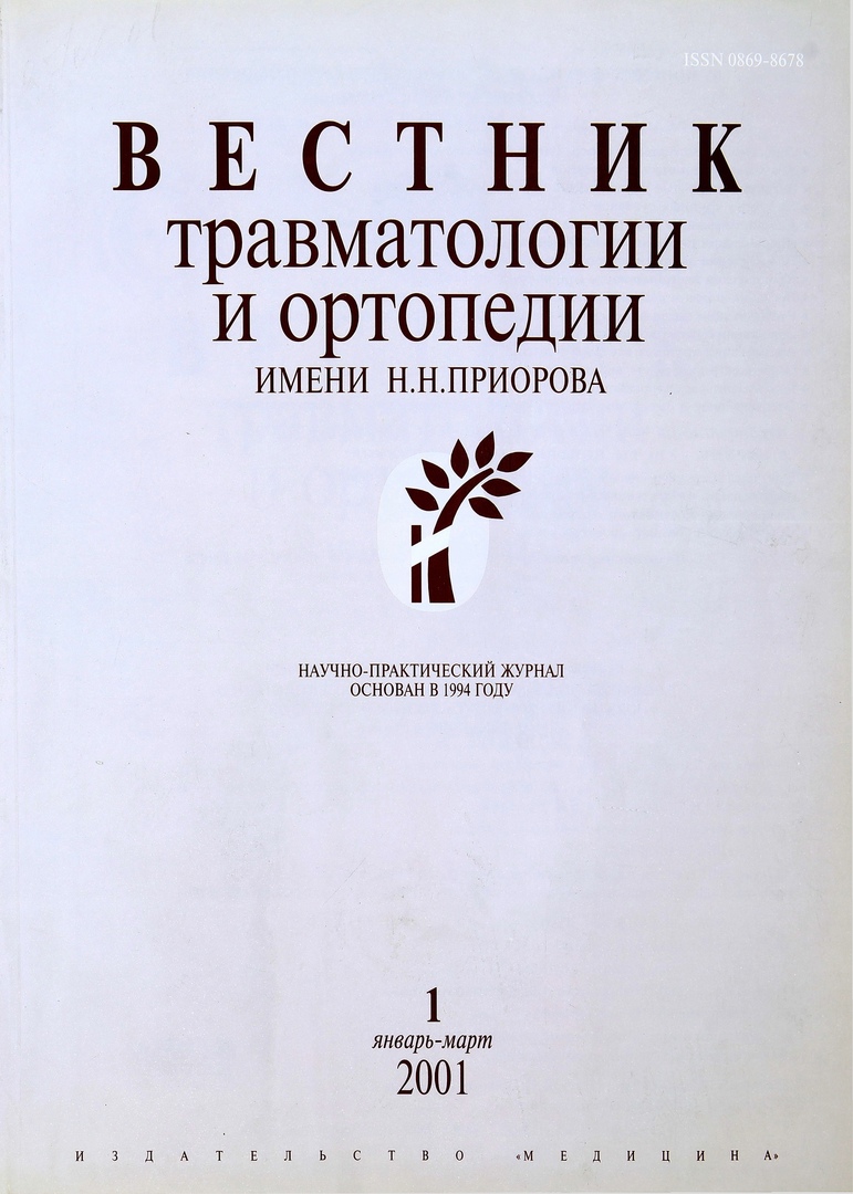
卷 8, 编号 1 (2001)
- 年: 2001
- ##issue.datePublished##: 02.03.2001
- 文章: 20
- URL: https://journals.eco-vector.com/0869-8678/issue/view/4914
- DOI: https://doi.org/10.17816/vto.01
完整期次
SCIENTIFIC REVIEWS
Massive bleeding in pelvic injuries: what to do?
摘要
Massive bleeding is the main cause of death in patients with extensive severe fractures and dislocations of the pelvic bones in the first minutes and hours after admission to the hospital [7, 45, 63]. In 54~88% of cases, the source of bleeding is damaged pelvic vessels [43, 63, 82]. In particularly severe pelvic injuries with predominant damage to its main blood vessels, mortality reaches 89-100% [43, 79].
 66-73
66-73


Lectures
Pathophysiological mechanisms of the consequences of trauma of the musculoskeletal system
摘要
In the most general form, the main factors of traumatic disease can be represented as follows (Scheme 1): the traumatic factor acts on organs and tissues, causing their damage. As a result of this, the cells are destroyed and their contents are released into the intercellular environment; other cells are exposed to concussion, as a result of which their metabolism and their inherent functions are disturbed. Primarily (as a result of the action of a traumatic factor) and secondarily (due to a change in the tissue environment), numerous receptors in the wound are irritated, which is subjectively perceived as pain, and objectively characterized by numerous reactions of organs and systems.
 62-65
62-65


Articles
Continuous improvement of the quality of medical care is the main direction of work of Russian orthopedic traumatologists (Part 2)*
摘要
Present work is a sequel to the publication presented in № 3, 2000. Projecting of the medical technological process is considered on the example of the hospital treatment of patients with low back degenerative diseases. Technologic chart for diagnosis and treatment of patients with spondylogenous low back pain for the inpatient treatment is given. High efficacy of the proposed technological approach to the treatment of lumbar spine degenerative diseases is proved by clinical results.
 3-10
3-10


Spinal osteomyelitis
摘要
Experience in diagnosis and treatment of 112 patients with spine osteomyelitis is presented. In 8 patients cervical spine, in 23 patients thoracic spine and in 81 patients lumbar spine was involved. Neurological deficit was observed in 45 (40.2%) patients. Forty seven (42%) patients underwent conservative and 65(58%) surgical treatment. Conservative treatment included intraarterial injection of antibacterial drugs. Surgical treatment consisted of radical resection of the osteomyelitis focus followed by stabilization of the spine with autografts. Long-term results were evaluated in terms from 1 to 20 years. In all patients the formation of bone block and regression of neurological symptoms was observed.
 11-16
11-16


Modern prevention and treatment of thrombotic complications in patients with polytrauma in the postresuscitation period
摘要
Results of examination and treatment of patients with polytrauma complicated by deep vein thrombosis of lower extrimities (DVT) and pulmonary thromboembolism (PTE) are presented. Proximal deep vein thrombosis was confirmed by Doppler. PTE developed on week 2 after trauma in patients with asymptomatic DVT. For timely diagnosis of DVT and prevention of PTE in patients with polytrauma it is necessary to perform Doppler of lower extrimities 1—2 times a week within 2—4 weeks after trauma and for 2—4 weeks after surgical interventions on long bones and pelvic bones. In patients with PTE the implantation cava-filter allowed to avoid lethal outcomes and PTE relapse. The application of low molecular weight heparin allowed to increase the efficacy ofprevention and treatment of thrombotic complications in patients with polytrauma.
 16-20
16-20


Therapeutic and diagnostic arthroscopy of the hip joint (first experience)
摘要
The experience in therapeutic diagnostic hip arthroscopy is presented. Hip arthroscopy was performed in 11 patients, aged 3-11 years. There were 5 patients with congenital hip joint dislocation, 3 patients with Legg-Kalve-Perthes disease, 2 patients with deforming coxarthrosis and 1 patient with pathologic hip dislocation. The original technique for hip arthroscopy is suggested. High efficacy of arthroscopy for the diagnosis and treatment of intraarticular hip pathology is shown.
 21-23
21-23


Aseptic necrosis of the femoral head as a complication of its traumatic dislocation
摘要
The work is based on the analysis of examination and treatment results in traumatic hip dislocation (52 patients). The development of aseptic femoral head necrosis was detected in 11 patients at follow-up from 2 to 5 years. Algorithm of complex examination and treatment in traumatic hip dislocation has been elaborated for early and long-term periods after trauma. The group of high risk for aseptic femoral head necrosis development includes the patients with concomitant trauma, with posterior, obturatory and central dislocation. Rehabilitation is requiered to all patients with traumatic hip dislocation in the period from 2 to 5 years taking into account the peculiarities of hip injury. If the signs of posttraumatic aseptic femoral head necrosis are detected at early stages, then conservative treatment is indicated. In advanced process the operative treatment is indicated. Localization of necrosis area within femoral head, necrosis extension and articular surface congruence are of importance for the choice of adequate operative treatment. Roentgenologic signs of irreversibility of articular tissues destruction are the indication for total hip replacement.
 24-28
24-28


Dynamics of Sonographic and Morphological Changes in Baker's Cyst Formation
摘要
Comparative analysis of sonographic and morphologic assessment in formation of Baker's cyst has been performed. There were 25 patients with gonarthrosis lasted 5.6 + years. In development of Baker’s cyst 3 periods were defined. Each period is characterized by structural changes caused by cyst development age and gonarthrosis stage. The period has specific quantitative and qualitative patterns of cyst wall and content. Ultrasound diagnosis of cyst structural development was reliably verified by histologic data. Ultrasound assessment of synovial masses in popliteal area allows to differentiate Baker’s cyst with other pathologic processes in deforming knee arthrosis of different genesis. Accurate definition of structural cyst development periods is useful for the detection of indications for cyst extirpation.
 29-32
29-32


Normal ultrasonographic picture of the tendons of the hand
摘要
The potentialities of ultrasonography for the imaging of wrist tendons is shown. Normal sonographic appearance of flexor wrist tendons as well as extensor wrist tendons is given in longitudinal and transverse scanning. The causes of artifacts have been analysed and the methods for their elimination are given. Elaborated protocol and technique of ultrasonographic investigation of wrist tendon at rest and in dynamics under real time condition are presented.
 33-36
33-36


Biomechanical substantiation of reconstructive surgery on the tendon of the long flexor of the first finger
摘要
The description of the technique and biomechanical grounds of the reconstructive operation (elaborated by the author) on the ten. flexor pollicis longum are presented. The operation is performed to eliminate hyperextension-flexion deformity of the thumb in old injuries of the ulnar nerve.
 36-37
36-37


Isolated avulsion fracture of the tibia at the insertion to the posterior cruciate ligament
摘要
Three cases of evulsion tibia fractures at the posterior cruciate ligament attachment are presented. The bone fragment displacement was more than 1 cm. All patients were treated surgically. In 1 patient reduction offracture was completed by bone suture, in 2 patients by fixation with screw inserted retrogradely. Follow up from 6 months to 4 years showed no complications, the joint was stable and full volume of motion was noted.
 38-40
38-40


Treatment of limb fractures in children with multiple and concomitant injuries
摘要
Experience in treatment of bone fractures in 105 patients with multiple and concomitant traumata is presented. There were 228 limb bones fractures. In 64 patients (61 %) conservative treatment including closed reposition, plaster immobolization, skeletal traction was used. Forty one patients (39%) were treated surgically, mainly by extrafocal osteosynthesis with external rod fixatives elaborated in our clinic. In the majority of patients (72%)) osteosynthesis was performed within the period from 4 to 7 days after trauma. In patients with crus and femur fractures the surgical treatment efficacy (by 100 points scale) for femur fractures was 37.9%), for crus fractures - 48.2%). In cases of combination of shoulder and forearm fractures the surgical efficacy treatment was 41.8%o and 42.3%o, respectively. In patients with unilateral femur and crus fractures the efficacy of treatment using rod fixatives made up 47.2%o. Efficacy was higher in all cases of extrafocal osteosynthesis as compared with traditional methods of treatment.
 40-43
40-43


Biomechanical Characteristics of Walking in Patients with Different Forms of Infantile Cerebral Palsy during Treatment with Dynamic Proprioceptive Correction
摘要
Results of biomechanic examination of walking in healthy individuals and patients with ICP before and after treatment using dynamic proprioceptive correction are presented. The patients with ICP had different forms of the disease: spastic, hemiplegic, hyperkinetic and atonic-ataxic. The following features of walking were studied: range and volume of flexor-extensor motion in hip, knee and ankle joints as well as time characteristics of step cycle. Data obtained showed common and individual signs of gait disturbance depending on the form of ICP. In author's opinion the possible mechanism of walking disturbance in patients with ICP was pathological change of motion stereotype and that disturbed motion stereotype was normalized using dynamic proprioceptive correction. Biomechanic examination of walking allows to provide the differentiation of various forms of ICP as well as to evaluate the locomotor function changes in the course of treatment.
 44-50
44-50


The use of electrochemically activated solutions in the traumatology and orthopedic clinic
摘要
During some years at clinical departments and laboratories of CITO electrochemically activated solutions are used for disinfection and sterilization. These solutions are synthesized in apparatus STEL—10N—120—1 (katalytic neutral anolyte). Results obtained are promising for the prevention of intrahospital infections. Practically no other disinfectants are used at the hospital.
 50-52
50-52


Use of Demineralized Bone Matrix to Repair Damaged Long Bones with Significant Defects
摘要
Introduction of tubular perforated implants of demineralized bone matrix was shown to provide the formation of organ specific bone regenerate and restoration of full value anatomic structure of the injured bone. Shape, material, method of matrix implantation conditioned the development of longitudinally oriented compact bone between free cortical distal and proximal bone fragments as well as the restoration of bone marrow intergrity in the defect site and its intergra-tion with bone marrow of total bone. Implant structure having multilevel widely branched canal system is suggested to provide free microcirculation of tissue liquid and migration of bone cell precursors into osteogenesis area.
 53-56
53-56


Embryonic development of the cruciate complex of the human knee joint: I. Anlage and primary differentiation
摘要
Twenty six human abortive embryons (6-13.5 weeks of prenatal development) were for the study of cruciate complex embryonal differentiation. Serial histological mounts (8—12 mkm) were prepared from leg fragments with knee joint. Mallory, hematoxylin-eosine and antibody for collagen III were used for staining. Human cruciate complex appears in embryons at 6-6.5 weeks of development. It differentiates from the mesenchyme lying between tibia and fibula. Starting from 8.5-9 weeks the anterior and double-component posterior ligaments, which begin to vascularize on the surface, can be distinguished. At the age of 13 weeks both components of posterior ligament fuse and the complex itself starts to vascularize throughout the whole mass.
 57-61
57-61


Irina Iosifovna Mirzoeva
摘要
In December 2000, Professor Irina Iosifovna Mirzoeva turned 80 years old. She was born on December 6, 1920 in a family of St. Petersburg lawyers. Her father I.A. Mirzoev and his stepfather - a well-known lawyer and writer from the group of "Smenovekhites" A.V. Bobrishev-Pushkin on false charges were arrested and shot. Only many years later, both of them are fully rehabilitated.
 74-74
74-74


Yuri Ivanovich Pozdnikin
摘要
Director of the Scientific Research Children's Orthopedic Institute named after A.I. G.I. Turner (GUN) to Doctor of Medical Sciences, Professor Yu.I. Pozdnikin.
 75-75
75-75


Nikolai Gavrilovich Fomichev
摘要
May 10, 2001 marks the 60th birthday and 36 years of medical and scientific and pedagogical activity of the Honored Doctor of the Russian Federation, Doctor of Medical Sciences, Professor N.G. Fomichev.
 76-76
76-76


Zagidulla Ismailovich Urazgildeev
摘要
January 1, 2001 marked the 60th anniversary of Z.I. Urazgildeev - Doctor of Medical Sciences, Professor, Head of the Department of the Consequences of Injuries of the Musculoskeletal System and Purulent Complications of the Central Research Institute of Traumatology and Orthopedics named after I.I. N.N. Priorov.
 77-77
77-77


