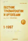
Vol 4, No 1 (1997)
- Year: 1997
- Published: 15.01.1997
- Articles: 21
- URL: https://journals.eco-vector.com/0869-8678/issue/view/5310
- DOI: https://doi.org/10.17816/vto.41
Full Issue
Original study articles
Modern realities in orthopedics and traumatology and prospects for the development of the specialty
Abstract
In historical terms, our specialty can be called both the oldest - medicine began with the treatment of injuries - and the young one, the beginnings of which appeared about two and a half hundred years ago, during the time of Nicolas Andry, but in fact - with the discovery of X-rays (1895).
 3-5
3-5


Treatment of bone loss defects and pseudoarthroses of the humerus with vascularised autografts
Abstract
Results of treatment of 31 patients with non-union of the humerus are presented. There are 25 defects of the humerus and 6 pseudoarthroses of various genesis and different localisation. Shortening of the humerus ranged from 3 to 15 cm. Routine free vascularised bone or composite skin/bone autografting was done for all patients. In 28 cases vascularised fibular pedicle graft was used, in 4 of which the fibular head to reconstruct the shoulder joint was used. An iliac crest graft was used in remaining 3. In 9 patients with shortening of the humerus over 5 cm distractionusing Ilizarov device was followed by bonee grafting. In 27 patients Ilizarov device was epplied for osteosynthesis after bone grafting. Long term outcome was studied in 27 of the 31 cases and the results were uniformly satisfactory.
 6-10
6-10


Transplantation of vascularized latissimus dorsalis flaps to the deltoid region for children with obstetrical brachial paralysis sequela
Abstract
Bipolar transplantation of vascularized pedicle flaps from the latissimus dorsalis to the deltoid region was done in 25 children between the ages of 2 and 14 years. All had sequela of obstetrical brachial plexus paralysis with various degrees of internal rotation contracture. Pre-operative limitation of active abduction ranged 0 to 80 degrees (mean=56 degrees). Muscle transplantation was supplemented in all cases by other musculotendinous and osteoarticular procedures. The surgical procedure and postoperative management are described in detail. All children showed improvement of arm function and shoulder range of motion. Post-operatively, active abduction was 20 to 170 degrees (mean=131 degrees). Internal rotation contracture was reduced and active external rotation was ebabled. There were two complications: one necrosis of the pedicle flap and one traction neuritis of the brachial plexus.
 10-15
10-15


Combination of craniocerebral and skeleton injurieis: diagnosis and management
Abstract
The study is concerned with the outcomes of the treatment of 364 patients with combined trauma (craniocerebral injuries of different severity and fractures of skeleton bones [453]). Patients’ management and fracture fixation methods are presented. Primary osteosynthesis (during the first day after trauma) was done in 45.9% of victims and delayed osteosynthesis - in 54.1%. The following fixators were applied: plates (46.6% of victims), external fixation devices (36.6%), rods (9.9%), other types (6.9%). Early osteosynthesis (during the first three days after trauma) did not increase the lethality and complication rate. At follow up period of 12-18 months the long term results were studied in 98 of 364 patients. Good outcome was achieved in 79.6% of cases, satisfactory - in 17.3% and unsatisfactory in 3.1% of cases. The authors consider that primary and early osteosynthesis in patients with the combination of craniocerebral and skeleton injuries enable to improve the outcomes, reduce the lethality rate, shorten the duration of hospitalisation and promote early rehabilitation.
 15-18
15-18


Complication rate of long bone fractures treated by early stable internal and trans-osseous osteosynthesis
Abstract
Complications occurring in 94 (24.1%) of 390 operative osteisynthesis were analysed. There were 284 patients with composite (115) and combined fractures of long bones. Complications occurred during recovery in 54 (23.6%) cases of transosseous osteosynthesis and in 40 (24.8%) cases of internal osteosynthesis. Intra-operative complications occurred in 4 cases due to technical errors, e.g. further comminution or vascular injury. Postoperative complications occurred in 7 cases due to inappropriate choice of fixation device, e.g. loss of reduction or fication failure. Postoperative infections predominantly involved the femur and tibia (60.6%). The final outcome was not influenced by local pin tract infection involving either skin, bone or both. There were 15 cases with general complications, e.g. pneumonia, thromboembolism, and decubiti; and 12 with local complications, e.g. toxidermia and marginal wound necrosis. All general complications were associated with restricted patient mobility and were observed 3 times more frequently (6.2%) with internal osteosynthesis than with transosseous osteosynthesis (2.2%). Careful attention to technique will help minimize the complication rate of osteosynthesis.
 18-23
18-23


Arthroscopic anterior cruciate ligament (ACL) reconstruction
Abstract
Arthroscopic «transtibial» ACL reconstruction has been frequently employed (38 cases) in our clinic practice since 1994. Details of the operative technique, special surgical instruments, post-opetative management and rehabilitation are described. Of all available grafts, we prefer free autografts from the middle third of the contralateral patellar ligament, with bone blocks at both ends (bone-tendon-bone). Our experience confirms the advatages of intracanal fixation with interference screws over the methods. Long term follow-up was available in 33 of the 38 patients, of which 32 had good or excellent results and one with an unsatisfactory outcome. This method can be recommended for the patient with an ACL deficient knee.
 23-27
23-27


Assessment of active stabilizators in capsular-ligamentous injuries of knee joint
Abstract
Clinical and instrumental testing technique of active stabilizators in capsular-ligamentous injuries of knee joint is presented. The score for the assessment of subjective and objective clinical signss of knee instability is shown in detail. It’s peculiarities consist of unification of these sings and their evaluation by 6-point scale. Test gives the integral index of the type form of knee instability. Protocols of knee joint function using «Biodex» are given. The testing is used to assess the function of periarticular muscles. Standard isokinetic test is used to examine the force and endurance, modified isometric test is used to evaluate the proprioception state and ability to response under different forces. Complex examination of 67 patients with various capsular-ligamentous injuries of knee joint confirms the high reliability and trustworthiness of data obtained.
 27-32
27-32


Biomechanics of «Femur-Bliskunov’s distractor» system in various osteotomies
Abstract
Bimechanical models of force interaction in «femurBliskunov’s distractor» system, biomechanical basis of various osteotomies (transverse, oblique, oblique transverse, Z-shape, Z-shape oblique) quantitative evaluation of osteotomy firmness parameters are done. The majority of osteotomies was performed with special device on the side of bone canal. Thirteen years experience of lengthening of 213 femurs (187 patients) confirms the calculated parameters. The authors prefer the Z-shape oblique osteotomy.
 33-40
33-40


Treatment of slipped capital femoral epiphysis complicated by chondrolysis of the hip joint
Abstract
The experience of treatment of 38 patient (39 joints) with slipped capital femoral epiphysis complicated by chondrolysis of the hip joint is presented. Clinical and radiologic examinations, comparable scintigraphy with 99mTc (10 patients) and morphologic examination (11 joints) were used. Camparable evaluation of outcomes of conservative (24 patients) and operative (14 patients) tratment confirmed the advisability of early surgical treatment. Mobilized-decompression procedure both an, independent one and in combination with reconstructive osteotomy is the method of choice. The follow up period ranged from 1.5 to 6 years. In 10 patients long term results were good, in 3 patients -satisfactory and in 1 patient - unsatisfactory.
 40-43
40-43


Improvement of blood supply and structure of avascular spongy bone: experimental study
Abstract
Model of avascular necrosis of the femoral head was induced bilaterally in the 52 hips (26 dogs). Immediately intertrochanteric osteotomy was done on 18 hips and 17 hips had autogenous muscle-pedicle bone graft. Seventeen were kept as controls. Intraosseous pressure was measured, the vascular architecture was studied, and histologic examination of the femoral heads was done at 4, 8, and 12 weeks. The authors consider that intertrochanteric osteotomy gives highly favorable effect on the structure of avascular femoral head and intravenous administration of nitroglycerin solution improves the intraosseous circulation.
 43-46
43-46


Lymphocytic dehydrogenases and immunologic reactivity in children with locomotor system injuries
Abstract
Immunologic reactivity was evaluated in 141 children between the ages of 6 and 15 with acute locomotor system trauma and its sequela. In 107 cases there were inflammatory complications of varying degrees in post-traumatic/ operative periods. Dynamics of succinic and a-glycerophosphate dehydrogenases was atudied, along with other parameters of immune competence. Inflammatory complications were associated with a decrease in lymphocytic dehydrogenase activity. There was also a correlation between the indices of immune reactivity and the status of the lymphocytic dehydrogenase enzymes. Metabolic medication maintained dehydrogenase activity and decreased the risk of post-traumatic and post-operative inflammatory complications.
 47-52
47-52


Local immunosupression as a factor for self-support of pathologic focus
Abstract
Cellular and humoral factors of immunosupression are largely the derivatives from injuried tissue. Polytrauma is proposed to be accompanied by the changes making special conditions for organism resistance. It is manifested in the induction of cellular supressive reactions and direct synthesis of autoimmune blocking humoral polypeptides and cytokines. Methods of sedative, antiblocking immunotherapy should be elaborated for successful treatment of infectious posttraumatic complications.
 52-56
52-56


From Practical Experience
Fixation of the endoprosthesis stem with muracito cortical bone allograft
Abstract
Endoprosthesis replacement of the femoral head for neck fractures in elderly and senile patients is the method of choice [1]. However, when using it, a number of significant factors should be taken into account. Bone tissue in this category of patients, as a rule, is porotic and characterized by increased fragility, the medullary canal is significantly expanded. Therefore, after the operation and subsequently, loosening of the endoprosthesis often occurs with all the ensuing consequences, such as pain, swelling of the limb, instability in the hip joint, inflammation, paraarticular calcifications. To ensure stable fixation of the endoprosthesis stem in the medullary canal of the femur, special cement, fragmented spongy auto- or allogeneic bone, hydroxyapatite glue, etc. are used [2–6]. However, these measures do not exclude the occurrence of a suppurative process, the development of micromobility, loosening and instability of the endoprosthesis.
 57-58
57-58


Method for the treatment of ulnar and prepatellar bursitis
Abstract
From 1992 to 1994, 118 patients with bursitis were treated in polyclinic No. 1 of the medical unit of the Ulyanovsk Automobile Plant - 111 (94.1%) with ulnar and 7 (5.9%) with prepatellar bursitis. All patients were male, aged 26 to 45 years. Serous bursitis occurred in 84 (71.2%) patients, and purulent bursitis in 34 (28.8%) patients. Trauma as the cause of bursitis was found in 83 patients. The main clinical sign of bursitis was the appearance of a soft, painless swelling on the dorsum of the elbow or anterior surface of the knee joint. Suppuration was manifested by redness, swelling, pain, fever. The treatment was carried out according to the generally accepted method (Struchkov V.I. et al., 1991). So, the final method of treatment in 71 patients was the puncture of the bag. At the same time, its contents were removed, 1-2 ml of 70° alcohol was injected, after which the area of the bag was massaged and the remaining alcohol was aspirated. In 6 patients, punctures had no effect and bursitis was regarded as chronic, and therefore bursectomy was undertaken. With purulent bursitis in all 34 patients, the purulent cavity was opened and drained.
 58-59
58-59


Arthroscopy of the knee joint (according to the materials of the traumatology department of the medical and sanitary unit of Zheleznogorsk)
Abstract
During the period from April 1992 to 1994 inclusive, operations using arthroscopic technique were performed on 50 patients in the traumatology department of the medical unit of Zheleznogorsk. Diagnostic and operative arthroscopy was performed using the Karl Storz device in the operating room, in full compliance with the rules of asepsis and antisepsis. According to the indications, spinal anesthesia, intravenous or (in some cases) endotracheal anesthesia were used.
 59-60
59-60


The case of surgical treatment of a victim with an injury to the common carotid artery
Abstract
Diagnosis of vascular injuries is sometimes very difficult. You should always remember about the possibility of such injuries, taking into account: the location of the wound (N.I. Pirogov also pointed out the value of this diagnostic feature), data on bleeding after injury, the degree of developed anemia, the presence, absence or weakening of the pulsation of the artery distal to the injury site , noise phenomena during auscultation, the presence or absence of a hematoma in the wound area, pain and paresthesia on the side of the wound, as well as ischemic contractures. The main danger in vascular injuries is bleeding, the intensity of which depends on the size of the wound and the defect in the vessel wall. The most dangerous are wide ruptures of large vessels, which include the common carotid artery.
 61-61
61-61


SCIENTIFIC REVIEWS
Anomalies of development and dysplasia of the upper cervical spine (clinic, diagnosis and treatment)
Abstract
As you know, the craniovertebral region - the place where the spine passes into the skull - includes the two upper cervical vertebrae (atlas and axis) and the basal part of the occipital bone. Between these bone formations, two joints (head joints) are formed. The lower joint of the head (atlantoaxial) provides rotational movements, while the upper joint (atlantooccipital) provides mainly flexion-extension [5]. Both joints are characterized by significant anatomical variations, congenital anatomical disorders are not uncommon, as well as injuries and osteoarticular diseases [3, 10].
 62-67
62-67


Anniversary
S. M. Zhuravlev
Abstract
On February 1, 1997, Doctor of Medical Sciences Professor Sergei Mikhailovich Zhuravlev celebrated his 60th birthday. His many years of scientific and practical work to improve the organization of traumatological and orthopedic care for the population of the country deservedly received wide recognition from the scientific community and practical healthcare workers. At the heart of this activity at all its stages lay a deep diversified study of injuries and orthopedic morbidity, the search for the most effective ways to prevent them and reduce adverse social consequences - temporary and permanent disability, mortality.
 68-68
68-68


Information
 69-69
69-69


Obituary
A. I. Bliskunov
Abstract
Alexander Ivanovich Bliskunov, Doctor of Medical Sciences, Professor, Head of the Department of Traumatology and Orthopedics of the Crimean Medical University, an outstanding doctor who occupied one of the first places in the world ranking of traumatologists and orthopedists, passed away.
 70-71
70-71


 72-72
72-72


